


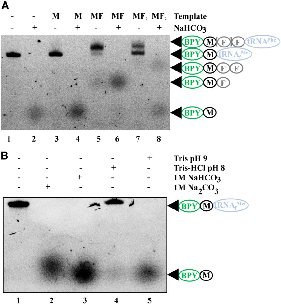
FIGURE 2.
Peptides attached to tRNA could be distinguished by urea PAGE following hydrolysis by 1 M NaHCO3. (A) Intact BPY-M-tRNAfMet (1) and products of M, MF, and MF2—coding mRNA translation, BPY-M-tRNAfMet (3), BPY-MF-tRNAPhe (5), BPY-MF2-tRNAPhe (7), and the products of their hydrolysis by 1 M NaHCO3 (2, 4, 6, 8) separated by urea PAGE. (B) Testing the conditions for BPY-M-tRNAfMet hydrolysis. Lanes correspond to the lack of treatment (1) and treatment with 1 M Na2CO3 (2), 1 M NaHCO3 (3), Tris-HCl (pH 8.0) (4), and Tris (pH 9.0) (5) as indicated. BPY is marked with a green oval—BODIPY label; black circle—methionine; blue oval—tRNAfMet or tRNAPhe.




