


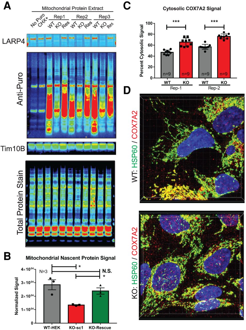
LARP4 promotes mitochondria-associated translation. (A,B) Analysis of mitochondria-associated translation rates by isolating mitochondria from puromycin-treated cells and measuring puromycin incorporation. (A) Puromycin immunoblots used for mitochondrial puromycin incorporation assay. (B) Quantification of average normalized mitochondria-associated puromycin signal from three independent biological replicates (N = 3). Controls include immunoblots for LARP4 to show depletion and TIM10B to show mitochondrial enrichment as well as a quantitative total protein stain for normalization. (C,D) Analysis of protein localization of COX7A2 (LARP4 target) by immunofluorescence staining with the mitochondrial protein HSP60 used as a mitochondrial marker and low threshold mask for cell area for image analysis. (C) Average percent cytosolic COX7A2 signal was quantified from nine fields of view (n = 9) of the HEK293LARP4−/− KO cells and WT cells from two independent biological replicates (Rep-1 and Rep-2). (D) Representative images from each condition and replicate are shown. Statistical significance of differences was assessed by two-tailed unpaired Student's t-tests (*) P ≤ 0.05, (***) P ≤ 0.0001. (N.S.) Not significant.




