


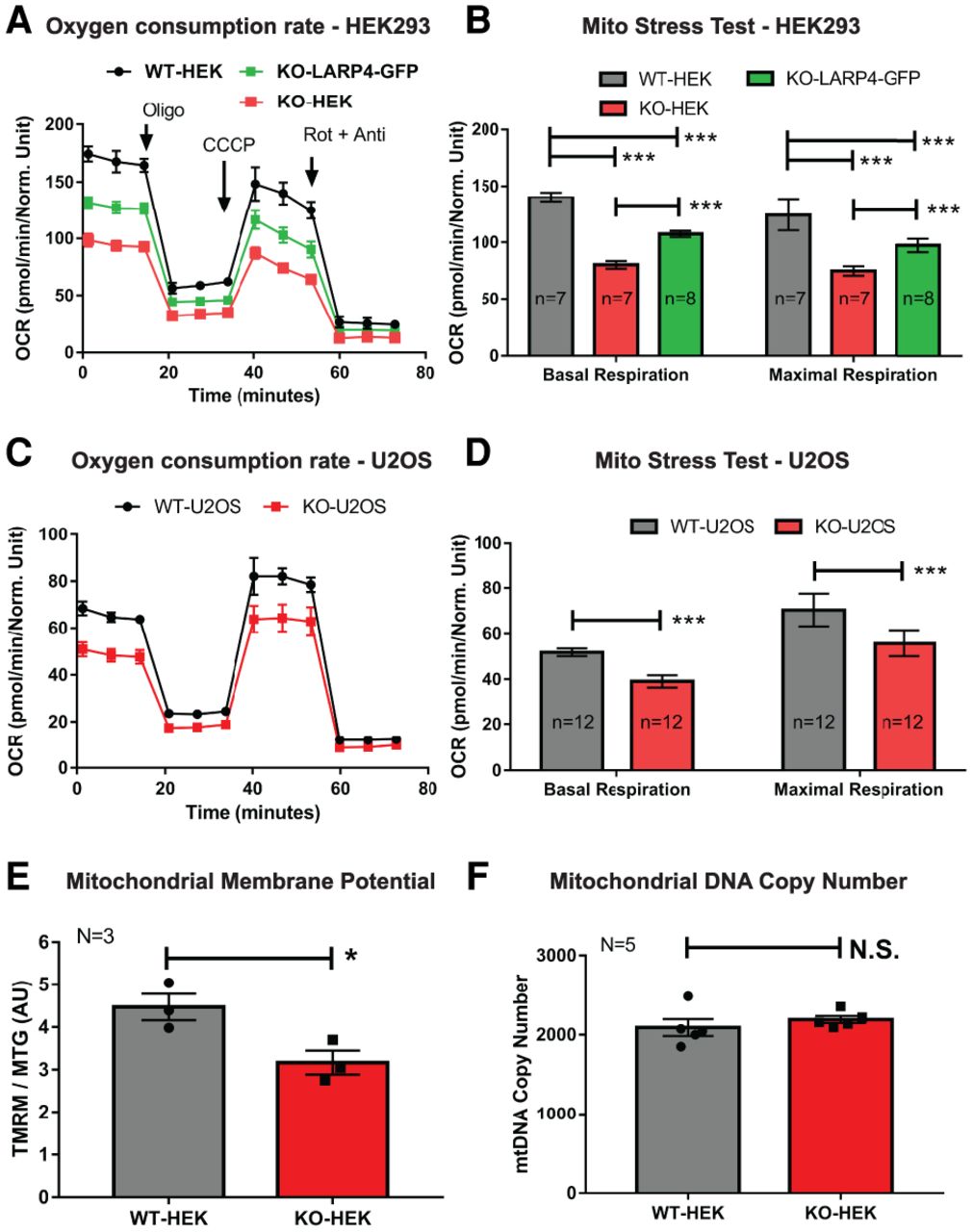
LARP4 promotes OXPHOS function. (A,B) Seahorse extracellular flux analysis of OCRs from WT HEK293 cells, HEK293LARP4−/− KO cells, and HEK293LARP4−/− KO-LARP4-GFP Rescue cells. Basal and maximal respiration rates (B) are calculated from the changes in OCR (A) in response to inhibitor addition. Averages shown are from technical replicates (N = 7 or 8, as indicated). (C,D) Seahorse extracellular flux analysis of OCRs from WT U2OS cells and U2OSLARP4−/− KO cells. Averages shown are from technical replicates (n = 12). (E) Analysis of mitochondrial membrane potential (MMP) by flow cytometry of WT HEK293 cells and HEK293LARP4−/− KO cells stained with the potential dependent dye tetramethylrhodamine–methyl-ester–perchlorate (TMRM) and the potential independent dye MitoTracker Green. Averages shown are from three independent experiments (N = 3). (F) Analysis of mitochondrial mass by proxy using qPCR measurements of mitochondrial DNA copy number from WT HEK293 cells and HEK293LARP4−/− KO cell samples. Averages shown are from samples from five independent experiments (N = 5). Statistical significance of differences was assessed by two-tailed unpaired Student's t-tests (*) P ≤ 0.05, (***) P ≤ 0.0001. (N.S.) Not significant. See also Supplemental Figure S4.




