


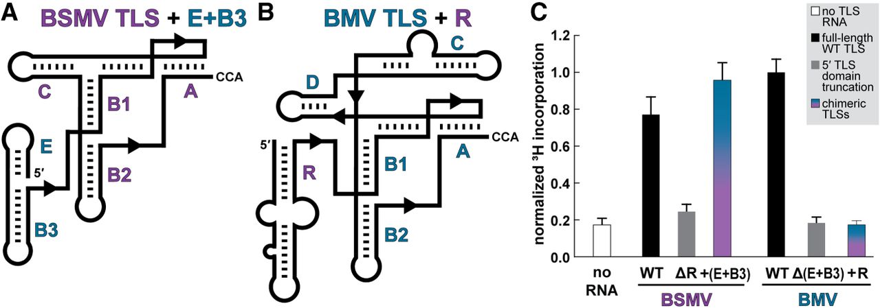
FIGURE 3.
Aminoacylation of chimeric TLSTyrs. (A, B) Cartoon diagrams of the secondary structure of the TLSTyr from (A) BSMV with the E and B3 stem–loops of BMV appended to the 5′ end in lieu of the R domain and (B) BMV with the R stem–loop of BSMV appended to the 5′ end in lieu of the E + B3 domain. (C) Relative activity of TyrRS on WT, truncated (ΔR or Δ[E + B3], gray), and chimeric (+[E + B3] or + R, blue-purple) TLSTyr RNAs as measured by 3H-tyrosine incorporation. See legend to Figure 2 for additional details.




