


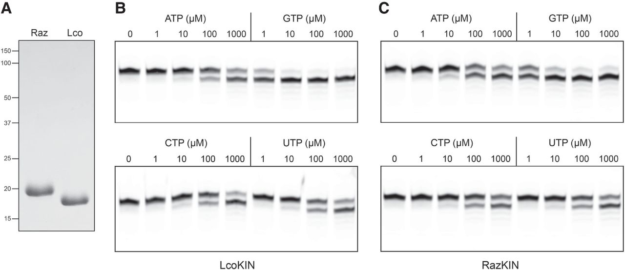
FIGURE 8.
RNA kinase activity of Rhizopus and Lichtheimia KIN proteins. (A) Aliquots (10 µg) of purified recombinant RazKIN and LcoKIN proteins were analyzed by SDS-PAGE. The Coomassie blue-stained gel is shown. The positions and sizes (kDa) of marker polypeptides are indicated on the left. (B,C) Reaction mixtures (10 µL) containing 50 mM Tris-HCl, pH 7.5, 50 mM NaCl, 2 mM DTT, 1 mM MgCl2, 100 nM (1 pmol) 10-mer 5′-OH RNA substrate 3′-labeled with Cy5, 5 nM (50 fmol) LcoKIN (B) or RazKIN (C), and 0, 1, 10, 100, or 1000 µM of the indicated NTP were incubated at 37˚C for 5 min. The reactions were quenched with formamide/EDTA, and the products were analyzed by urea-PAGE.




