


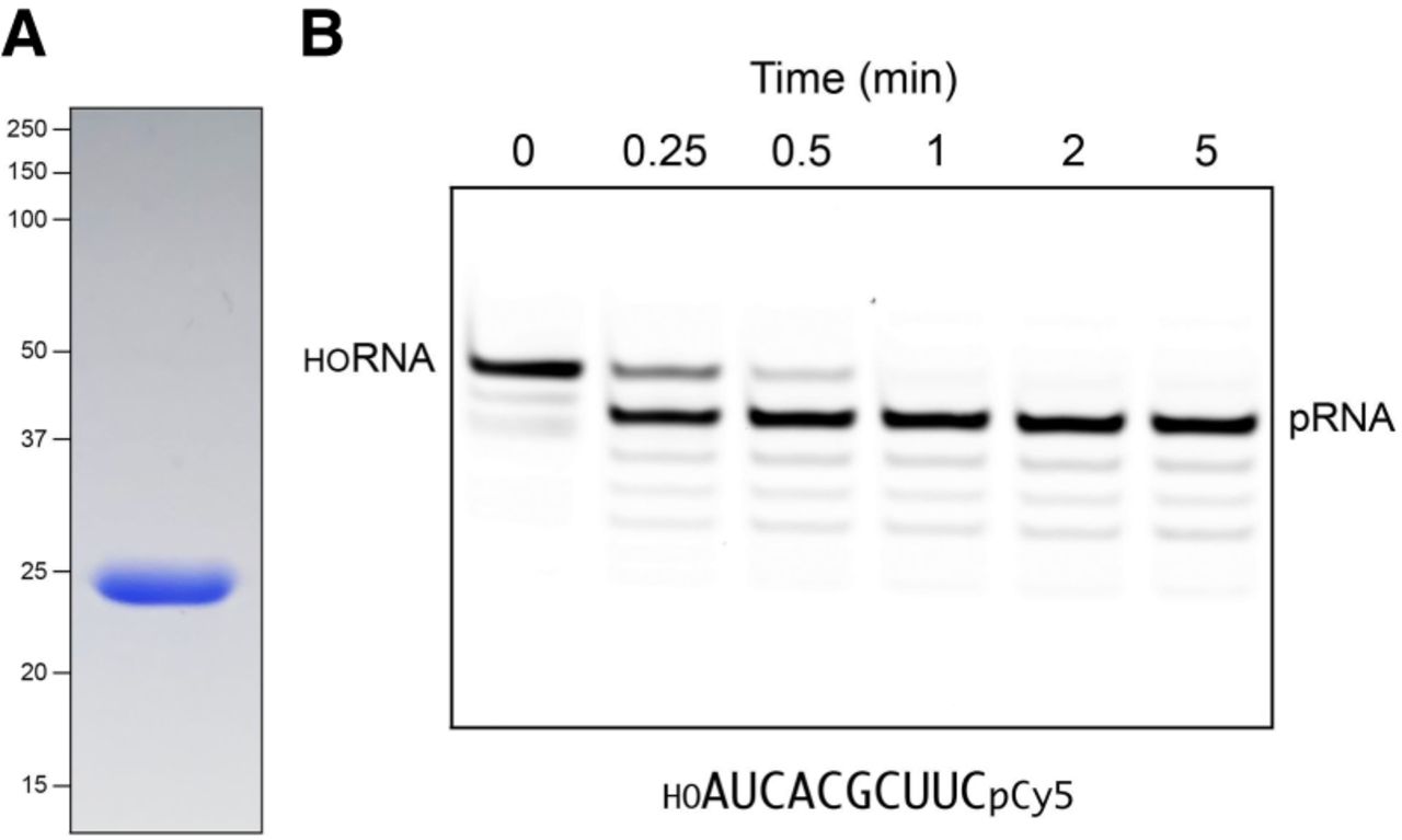
RNA kinase activity of recombinant MciKIN. (A) An aliquot (10 µg) of purified recombinant MciKIN was analyzed by SDS-PAGE. The Coomassie blue-stained gel is shown. The positions and sizes (kDa) of marker polypeptides are indicated on the left. (B) A reaction mixture (110 µL) containing 50 mM Tris-HCl, pH 7.5, 50 mM NaCl, 2 mM DTT, 1 mM MgCl2, 0.1 mM GTP, 100 nM 10-mer 5′-OH RNA substrate 3′-labeled with Cy5 (depicted at bottom; purchased from IDT), and 5 nM MciKIN was incubated at 37˚C. The reactions were initiated by adding MciKIN to a prewarmed reaction mixture. Aliquots (10 µL) were withdrawn prior to adding enzyme (time 0) or at the times specified after enzyme addition and quenched immediately with an equal volume of 90% formamide, 50 mM EDTA. The products were analyzed by electrophoresis (at 10 W constant power) through a 15 cm 20% polyacrylamide gel containing 7 M urea in 45 mM Tris-borate, 1 mM EDTA. The Cy5-labeled RNAs were visualized by scanning the gel with a Typhoon biomolecular imager (Cytiva). The positions of the 10-mer HORNA substrate and the 10-mer pRNA product are indicated.




