


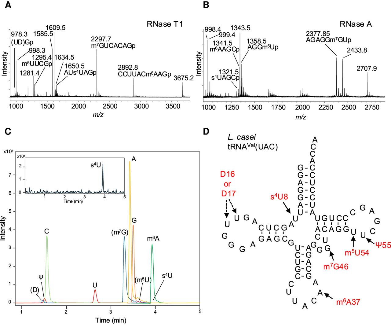
Mass spectrometry analysis of the native L. casei tRNAVal. (A) The MALDI-TOF mass spectrum of the native L. casei tRNAVal digested by RNase T1 shows s4U8 containing fragment AUs4UAGp, D containing fragment (UD)Gp, m6A37 containing fragment CCUUACm6AAGp, m7G46 containing fragment m7GUCACAGp, and m5U54 containing fragment m5UUCGp. (B) The MALDI-TOF mass spectrum of the native L. casei tRNAVal digested by RNase A shows s4U8, m6A37, m7G46, and m5U54. Supplemental Tables S1 and S2 give theoretical and empirical masses of singly protonated ions derived from the RNase T1 and RNase A fragments of the L. casei tRNAVal, respectively. (C) LC-HRMS analysis of nuclease P1 digested L. casei tRNAVal nucleosides. Overlay of chromatograms obtained for A, C, G, U, Ψ, (D), m6A, and s4U detected at m/z of 268.1040, 244.0928, 284.0989, 245.0768 (U and Ψ), 247.0925, 282.1197, and 261.0540, respectively, at the corresponding retention time expected from the authentic nucleoside samples. Ions at m/z of 259 and 298 are annotated as m5U and m7G, respectively. LC-HRMS data for s4U is provided in the inset. (D) Modified nucleosides of the native L. casei tRNAVal detected by mass spectrometry analyses. Detected modified nucleosides and their positions are shown in red letters.




