


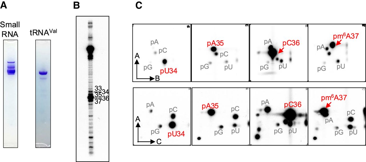
Nucleotide analysis of anticodon region in L. casei tRNAVal by Kuchino's post-labeling method. (A) L. casei tRNAVal was purified from the small RNA fraction. The purified small RNA and tRNAVal were analyzed by 10% PAGE (7 M urea), and the gels were stained with toluidine blue. (B) The purified tRNAVal was partially cleaved by formamide, and then the 5′-end of each fragment was labeled with γ-32P-ATP using T4 polynucleotide kinase. The RNA fragments were separated by 15% PAGE (7 M urea). Numbers correspond to the nucleotide positions in tRNAVal. (C) RNA fragments cleaved by formamide were purified from gel and digested with nuclease P1, and then their 5′ mononucleotides were analyzed by 2D TLC. The TLC analysis was performed in two types of solvent system, namely solvents A and B (upper four panels) or A and C (lower four panels). Solvent A was isobutyric acid:aqueous ammonia:water (66:1:33, v/v/v). Solvent B was isopropanol:HCl:water (70:15:15, v/v/v). Solvent C was 50 mM sodium phosphate, pH 6.8:ammonium sulphate:n-propanol (100:60:2, v/w/v). Nucleotides at positions 34–37 are shown. U34 was unmodified U, and spots of pm6A were detected at position 37.




