


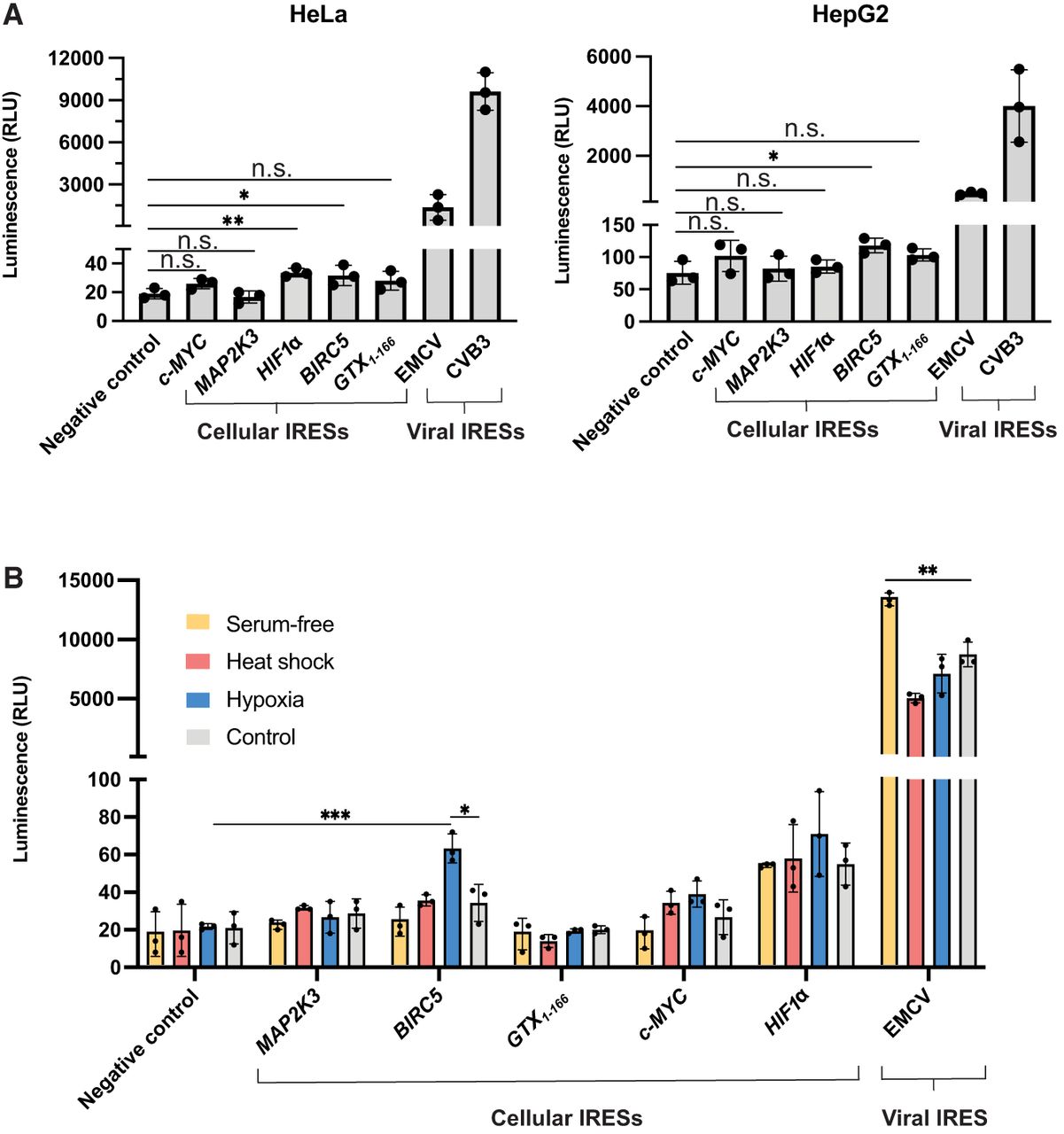
Circular split nLuc reporter can be used to test IRES activity in alternative cell types and states. (A) Luminescence from HeLa and HepG2 cells expressing the circular split nLuc reporter with a putative IRES. The overall trend in IRES activity of viral and cellular IRESs is similar in HepG2, HeLa, and HEK293T cells. (B) Luminescence from HEK293T cells transfected with the circular split nLuc reporter with a putative IRES. Cells were subjected to serum-free media (yellow), heat shock (salmon), hypoxia (blue), and no treatment control (gray). (RLU) Relative luminescence units. Data are presented as mean values +/− one SD (n = 3 biological replicates). Significance was calculated using unpaired two-tailed Student's t-test. (****) P < 0.0001, (***) P < 0.001, (**) P < 0.01, (*) P < 0.05, (n.s.) P > 0.05.




