


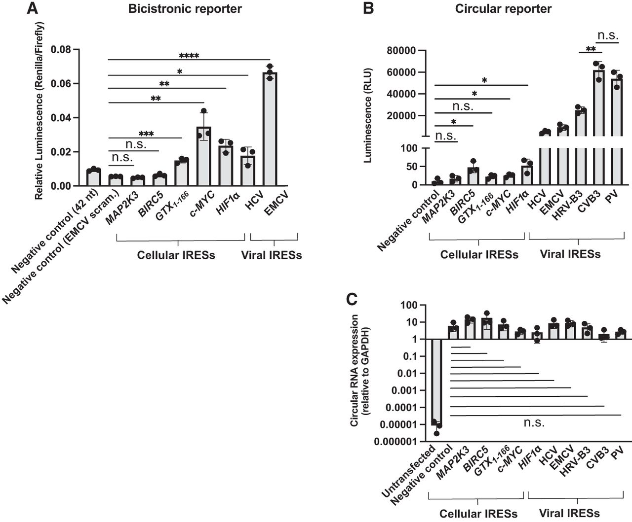
Circular split nLuc reporter accurately measures IRES activity. (A) Renilla luciferase normalized to firefly in HEK293T cells transfected with bicistronic reporters containing a putative IRES. Negative control is a scrambled EMCV sequence. (B) Luminescence from HEK293T cells expressing the circular split nLuc reporter with a putative IRES. (C) Circular RNA expression of the circular split nLuc reporter in HEK293T cells transfected with a putative IRES. Divergent primers that spanned the circularization junction were used for qPCR. RNA expression was normalized to GAPDH. The IRES does not affect circularization efficiency. (RLU) Relative luminescence units. Data are presented as mean values +/− one SD (n = 3 biological replicates). Significance was calculated using unpaired two-tailed Student's t-test. (****) P < 0.0001, (***) P < 0.001, (**) P < 0.01, (*) P < 0.05, (n.s.) P > 0.05.




