


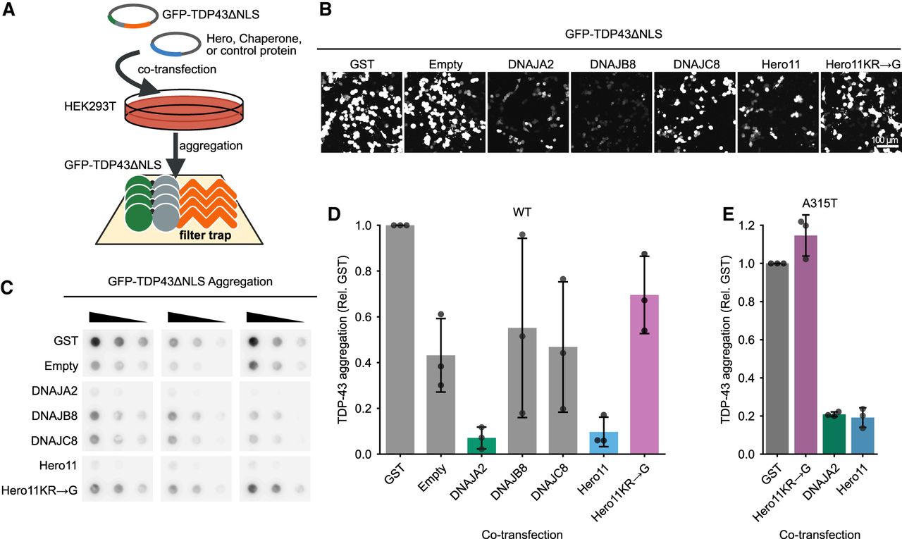
DNAJA2 and Hero11 suppress the aggregation of TDP-43 in HEK293T cells. (A) Schematic of the HEK293T cell-based filter trap assay for TDP-43 aggregation. Plasmids for GFP-TDP43ΔNLS and the protein indicated in (B) were cotransfected into HEK293T cells and incubated for 48 h. The cells were subsequently lysed and filtered through a 0.2 μm filter membrane under vacuum, on which aggregated proteins become trapped for detection via immunoblotting for GFP. (B) Representative microscope images of GFP-TDP43ΔNLS signals in HEK293T cells cotransfected with the indicated chaperones, Hero proteins or control at 20× optical magnification. Empty refers to the pCAGEN plasmid without any insert protein. (C) Immunoblots for aggregated GFP-TDP43ΔNLS in HEK293T cells detected with α-GFP antibody. For each filter trap membrane (from three independent experiments), each sample was loaded three times at twofold serial dilution. (D) Quantification of GFP-TDP43ΔNLS WT aggregation density in C. For each membrane, values are normalized to the aggregation in the GST cotransfection. Bar plots represent the mean and standard deviation as the bar height and error bar, respectively. Dots represent the values of individual experiments. (E) Quantification of GFP-TDP43ΔNLS A315T aggregation density normalized to aggregation in GST cotransfection for each membrane (from three independent experiments). Bar plots represent the mean and standard deviation as the bar height and error bars, respectively. Dots represent the values of individual experiments.




