


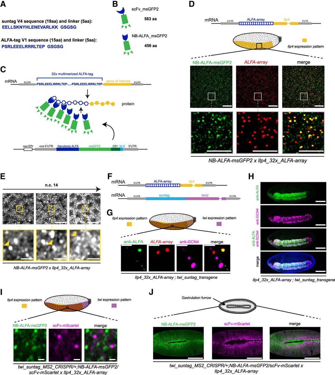
Development of the ALFA-array system to detect nascent translation in Drosophila. (A) Peptide sequence of one suntag with its linker (24 amino acids) and peptide sequence of the ALFA-tag with its linker (20 amino acids). (B) Schematic of the scFv and the nanobody ALFA (NB-ALFA) fused to the FP msGFP2 and their size in amino acids (aa). (C) Schematic of the ALFA-array system developed in this study to detect translation. Thirty-two ALFA-tag sequences were multimerized (blue amino acid sequence) and inserted at the 5′ of the gene of interest sequence (yellow). Upon translation, nanobodies ALFA fused to msGFP2 (NB-ALFA_msGFP2) will bind to the ALFA-tag peptides. As a bipartite system, the NB-ALFA is expressed under nos EPr fused to msGFP2, Streptococcal protein G (GB1) domain, and a nuclear localization signal (NLS). (D) Representation of the transgene mRNA containing 32x_ALFA-array in frame with the insulin-like peptide 4 (Ilp4) gene sequence. Schematic of a sagittal view of a Drosophila embryo with Ilp4 expression pattern in yellow. Anterior side is on the left, posterior on the right, dorsal on the top, and ventral side on the bottom. Black square represents the imaged area of the bottom panels. One Z-plane of confocal images from smFISH with direct Nb-ALFA-msGFP2 FP signal (green) and 32x_ALFA-array probes (red) on NB-ALFA_msGFP2 > Ilp4_32x_ALFA-array embryos in early-mid n.c. 14 is on the ventral side. Scale bars, 10 µm on the larger images, and 5 µm on the zoomed images. (E) Snapshots from representative fast-mode acquired confocal movies of Ilp4_32x_ALFA-array/+ n.c. 14 embryos carrying NB-ALFA_msGFP2. Bright white foci represent nascent translation of Ilp4 transgene. Yellow squares represent zoomed images of the top panels. Nascent translation of Ilp4 transgene is indicated by yellow arrowheads. Scale bars, 10 µm on the larger images, and 2 µm on the zoomed images (see related Supplemental Movie S24). (F) Representation of the transgene mRNAs containing 32x_ALFA-array fused to Ilp4 coding sequence or suntag fused to twist coding sequence used in G and H. (G) Schematic of a Drosophila embryo on the ventral side with twi expression pattern in purple and Ilp4 in yellow. Black square represents the imaged area of the bottom panels. One Z-plane of confocal images from immuno-smFISH with anti-ALFA (green) labeling nascent translation of Ilp4_32x_ALFA-array, probes against 32x_ALFA-array (red) labeling Ilp4_32x_ALFA-array mRNA molecules and anti-GCN4 antibody labeling nascent translation of twi_suntag_transgene (magenta) in n.c. 14 embryos. Scale bar, 1 µm. (H) Maximum intensity projection of a whole embryo confocal image from immunostaining with anti-ALFA antibody (green) and anti-GCN4 antibody (magenta) on Ilp4_32x_ALFA-array; twi_suntag_transgene gastrulating embryo. Scale bar, 100 µm. (I) Schematic of a Drosophila embryo on the ventral side with twi expression pattern in purple and Ilp4 in yellow. Black square represents the imaged area of the bottom panels. Single Z-plane of confocal images from dual-color live imaging of twi_suntag_MS2_CRISPR/+; NB-ALFA_msGFP2/scFv-mScarlet x Ilp4_32x_ALFA-array embryos in n.c. 14. Scale bar, 1 µm (see related Supplemental Movie S26). (J, top) Schematic of a Drosophila embryo on the ventral side with gastrulation furrow represented with invaginating cells. Confocal snapshots of a whole live embryo expressing twi_suntag_MS2_CRISPR/+; NB-ALFA_msGFP2/scFv-mScarlet x Ilp4_32x_ALFA-array, after gastrulation. Note the cytoplasmic staining from Ilp4 translation (green) and nuclear staining from twi translation (purple). Scale bar, 100 µm.




