


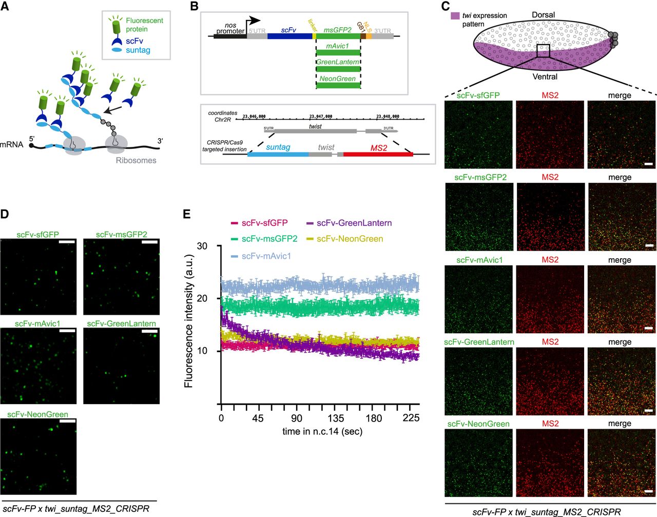
Generation of different scFv-fluorescent proteins (scFv-FPs) to monitor nascent translation in Drosophila. (A) Schematic of the SunTag system. Once the mRNA (black) is translated by the ribosomes (gray), the suntag epitopes (light blue) will be bound by the single-chain variable fragment (scFv) (dark blue) fused to an FP (green). Therefore, the nascent peptide will be detected by the accumulation of fluorescent signals. (B, top) Schematic of the different constructs of scFv-FP generated in this study. scFv-msGFP2, scFv-GreenLantern, scFv-mAvic1, and scFv-NeonGreen were created for this study, and scFv-sfGFP in our previous study (Dufourt et al. 2021). (Bottom) Schematic of the CRISPR/Cas9 targeted insertion strategy to obtain endogenous twist gene tagged with suntag and 128xMS2 (Dufourt et al. 2021). (C, top) Schematic of a sagittal view of a Drosophila embryo with twi expression pattern in purple. Anterior side is on the left, posterior on the right, dorsal on the top, and ventral side on the bottom. Black square represents the imaged area of the bottom panels. (Bottom) One Z-plane of confocal images from smFISH with direct FP signal in green (scFv-msGFP2, scFv-GreenLantern, scFv-mAvic1, scFv-NeonGreen, and scFv-sfGFP) and MS2 probes (red) on scFv-FP x twi_suntag_MS2_CRISPR embryos in early-mid n.c. 14. Scale bar, 5 µm. (D) Snapshots from representative fast-mode acquired confocal movies of twi_suntag_MS2_CRISPR/+ embryos carrying either scFv-msGFP2, scFv-GreenLantern, scFv-mAvic1, and scFv-NeonGreen or scFv-sfGFP proteins. Green dots represent nascent translation of twi. Scale bar, 5 µm (see related Supplemental Movies S8–S12). (E) Quantification across time during n.c. 14 of different scFv-FP signals from movies represented in D, n = 5 movies from at least three embryos for each scFv-FP. Error bars represent SEM.




