


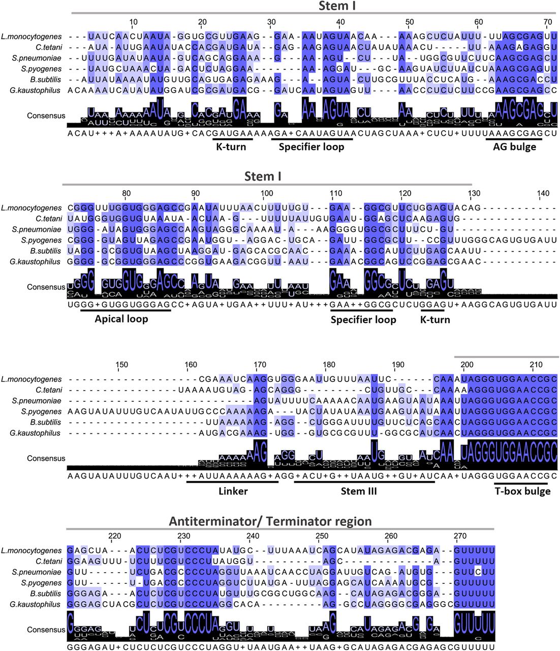
FIGURE 2.
Multiple sequence alignment of the glycyl T-boxes from L. monocytogenes, C. tetani, S. pneumoniae, S. pyogenes, B. subtilis, and G. kaustophilus. The alignment was performed using Clustal Omega, and Jalview 2.11.1.7 was used for illustration purposes. For the alignment, the highly conserved nucleotides (80%–100%) are indicated by dark blue color, the moderately conserved nucleotides (50%–80%) are indicated with blue, while the less conserved nucleotides (≤50%) are indicated with light blue. Alignment numbering does not correspond to the numbering of the respective glycyl T-boxes.




