


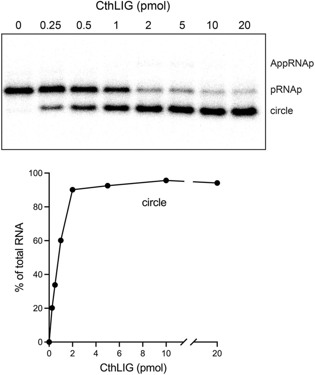
ATP-independent ligation by CthLIG–AMP. (Top panel) Reaction mixtures (10 µL) containing 50 mM Tris-HCl, pH 8.0, 2 mM DTT, 5 mM MgCl2, 1 pmol (0.1 µM) 32P-labeled 10-mer pRNA2’p, and CthLIG protein as specified were incubated at 37°C for 5 min and then quenched with formamide/EDTA. The mixtures were analyzed by urea-PAGE and the radiolabeled RNAs were visualized by scanning the gel. The positions of the pRNAp substrate, AppRNAp intermediate, and ligated circle product are indicated on the right. (Bottom panel) The extent of ligation is plotted as a function of input CthLIG. Each datum in the graph is the average of three independent titration experiments ± SEM. The error values are small, such that the error bars do not extent beyond the symbols.




