


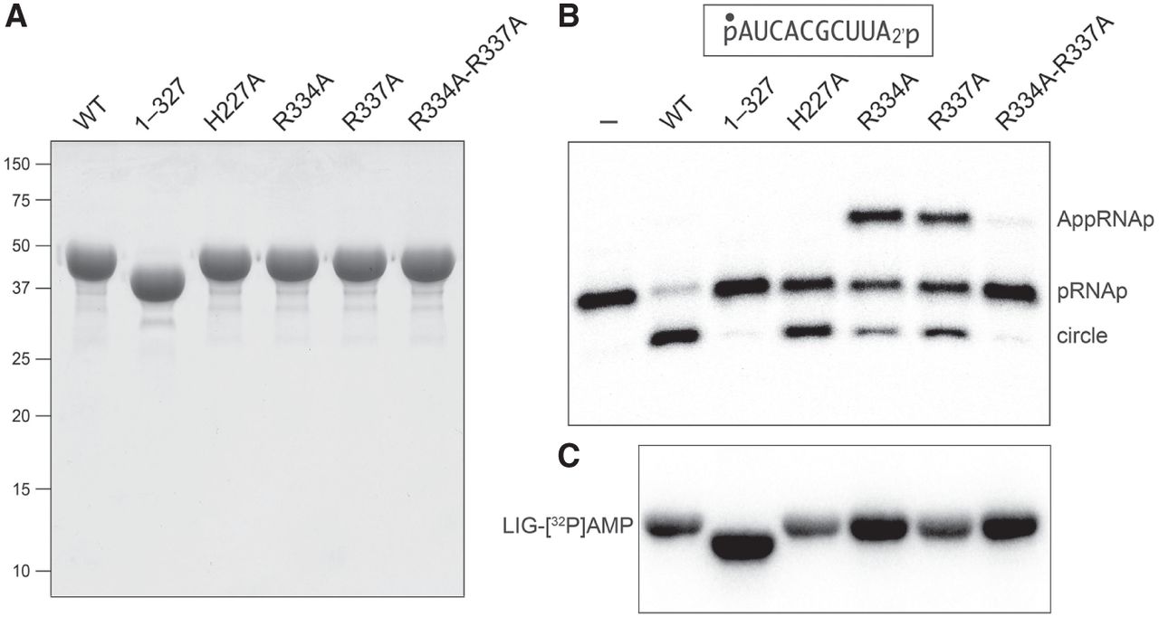
Structure-guided mutagenesis of CthLIG. (A) Aliquots (10 µg) of the purified full-length wild-type CthLIG, the indicated full-length alanine mutants, and the N-terminal adenylyltransferase domain (1–327) were analyzed by SDS-PAGE. The Coomassie blue-stained gel is shown. The positions and sizes (kDa) of marker polypeptides are indicated on the left. (B) Ligation reaction mixtures (10 µL) containing 50 mM Tris-HCl, pH 8.0, 50 mM NaCl, 2 mM DTT, 10 mM MgCl2, 100 µM ATP, 1 pmol (0.1 µM) 32P-labeled 10-mer pRNA2′p (shown at the top, with the 32P-label indicated by filled circle [•]), and either no enzyme (lane –), or 1 pmol (0.1 µM) of the indicated CthLIG protein were incubated at 37°C for 30 min. The reactions were quenched with an equal volume of 95% formamide/50 mM EDTA, and the products were analyzed by electrophoresis (at 58 W constant power) through a 40 cm 20% polyacrylamide gel containing 7 M urea in 45 mM Tris-borate, 1 mM EDTA. The radiolabeled RNAs were visualized by scanning the gel with a Typhoon FLA-7000 imaging device. The positions of the 5′-PO4,2′-PO4 RNA substrate (pRNAp), the 5-adenylylated intermediate (AppRNAp), and the 10-mer circle product of intramolecular ligation are indicated on the right. (C) Ligase adenylylation. Reaction mixtures (10 µL) containing 50 mM Tris-HCl, pH 8.0, 10 mM MgCl2, 50 µM [α32P]ATP, and 25 µM (250 pmol) of the indicated CthLIG protein were incubated at 37˚C for 5 min, then quenched with SDS, and analyzed by SDS-PAGE. A scan of the gel is shown.




