


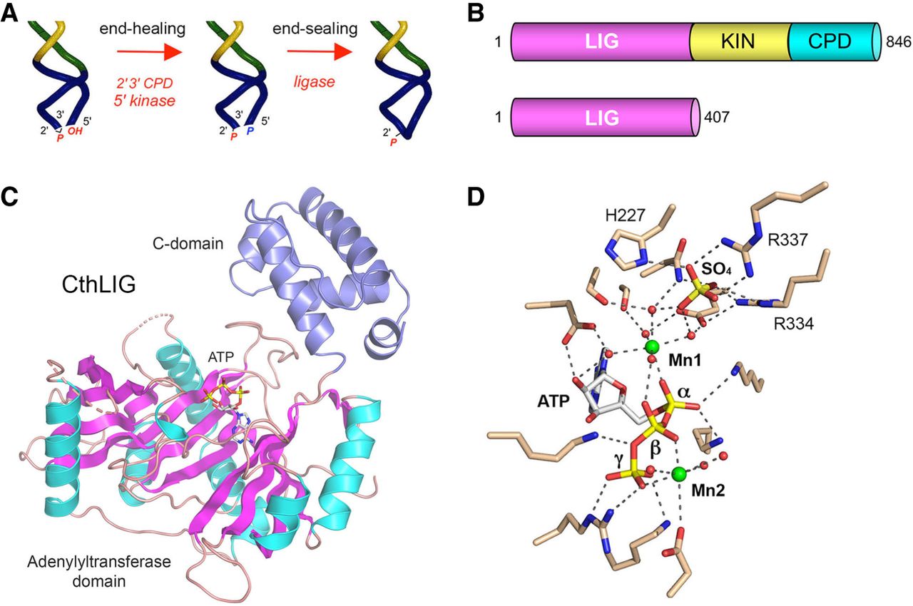
Domain structure and candidate 2′-PO4 binding site of Chaetomium thermophilum LIG (CthLIG). (A) Illustration of the end-healing and end-sealing phases of the fungal tRNA splicing pathway. (B) Cartoon depiction of the domain organization of full-length 846-aa C. thermophilum Trl1 consisting of N-terminal ligase (LIG), central kinase (KIN), and C-terminal cyclic phosphodiesterase (CPD) domains. The N-terminal 407-aa segment comprising an autonomous LIG enzyme is the subject of the present study. (C) The tertiary structure of CthLIG is shown with the N-terminal adenylyltransferase module colored by secondary structure (magenta β strands and cyan α helices) and the unique C-terminal domain (aa 328-406) colored blue. ATP bound to the adenylyltransferase domain is depicted as a stick model. (D) View of the CthLIG active site depicting atomic contacts to ATP, two Mn2+ ions (green spheres), and metal-bound waters (red spheres). The image highlights a sulfate anion (stick model) bound to the C-domain, adjacent to the catalytic metal (Mn1), that is thought to mimic the 2′-PO4 of the RNA substrate. Sulfate-binding residues His227, Arg334, and Arg337 were mutated to alanine in the present study.




