


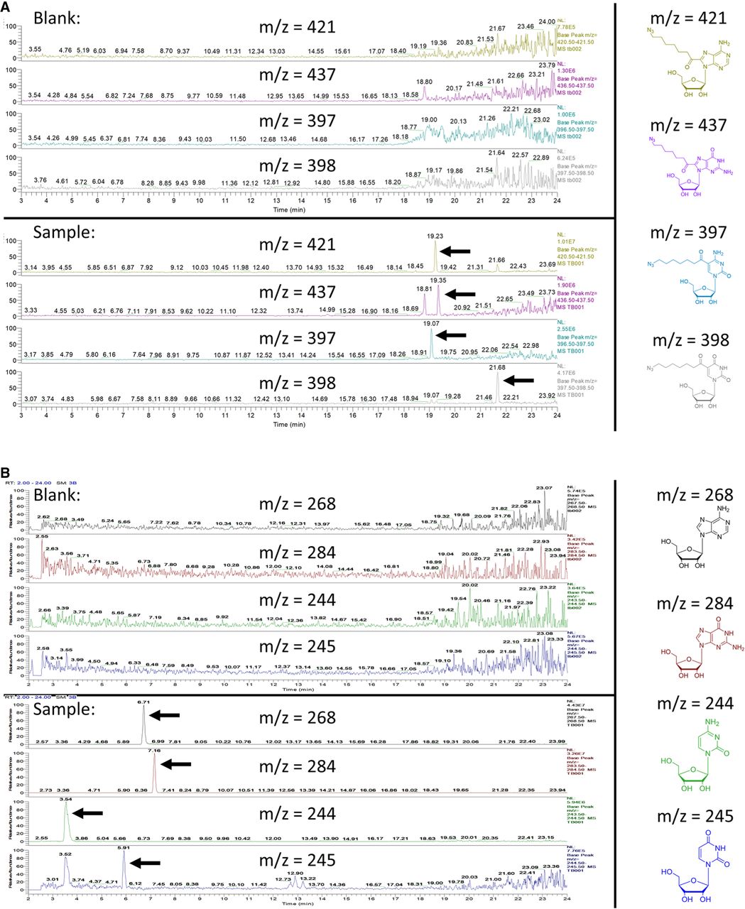
(A) LC/MS chromatograms of the four DAAS-labeled nucleosides (indicated by arrows), produced from a digested DAAS-labeled O.i. sample. A blank is included. Structures are included on the right, color coded to their respective chromatograms. The extra peak for DAAS-labeled guanosine at 18.81 min results from an unrelated mass from the blank. The peak at 19.35 min corresponds to the actual DAAS-labeled guanosine species. (B) LC/MS chromatograms of the four canonical nucleosides (indicated by arrows), produced from a digested DAAS-labeled O.i. sample. A blank is included. Structures are included on the right, color coded to their respective chromatograms. The extra uridine peak at 3.52 min results from cytidine overlap (likely caused by natural 13C incorporation) due to their difference of only 1 AMU.




