


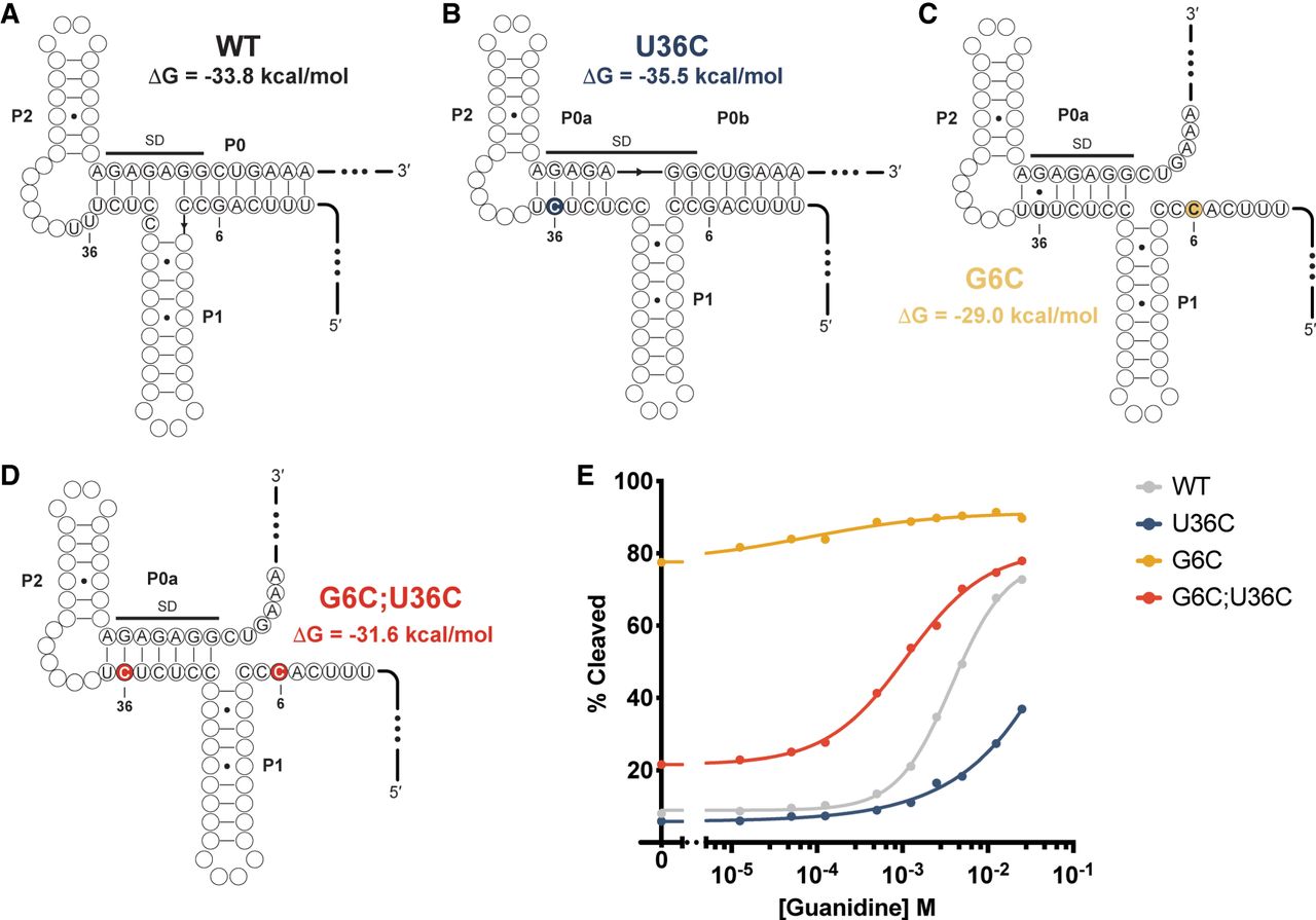
FIGURE 6.
Modeling the role of U36C in riboswitch function. (A) The WT MFE secondary structure of the Pae gdm-II riboswitch. Ribosome binding site shown in navy. (B) The U36C MFE secondary structure. U36C mutation is shown in yellow. (C) The G6C MFE secondary structure. G6C mutation shown in red. (D) The G6C;U36C MFE secondary structure. G6C and U36C mutations are shown as light blue. (E) Representative response profiles of the WT, G6C, U36C, and G6C;U36C Pae gdm-II riboswitches.




