


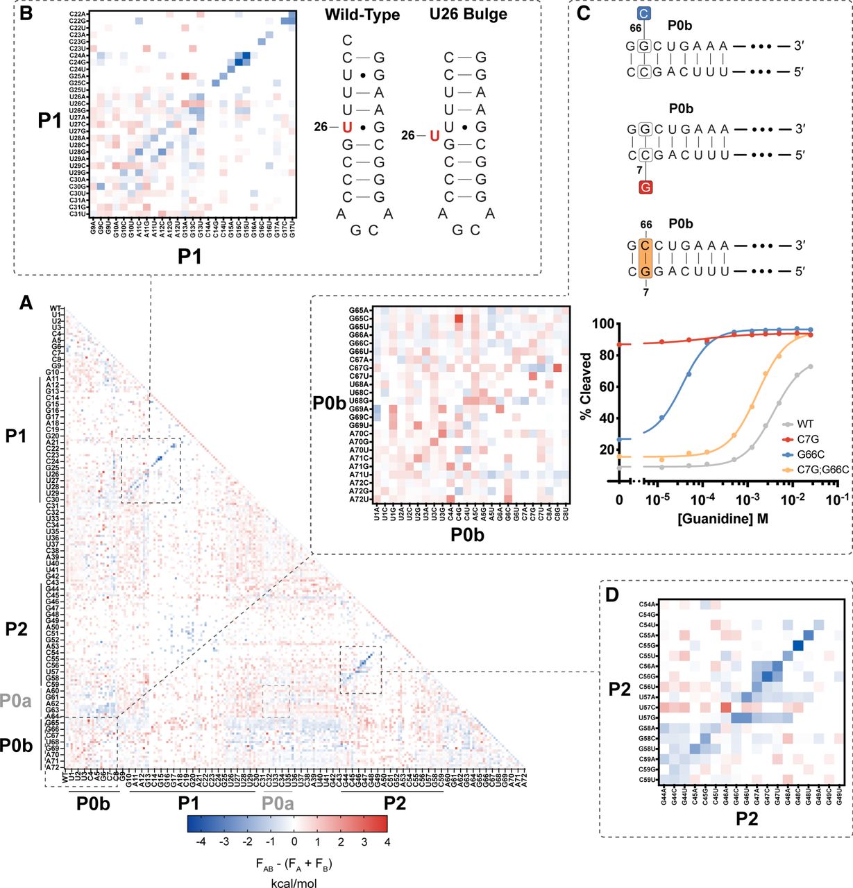
FIGURE 3.
Double mutant interactions reveal structural features. (A) Epistasis heat map of double mutant function parameters. Negative values reveal P1 and P2 helices, while positive values identify P0b. (B) Inset of P1 covariation with parallel diagonal indicating the U26 bulge shown in the accompanying secondary structure. (C) Inset of P0b covariation and secondary structure of key single mutations to a P0 base pair and the double mutant restoration. (D) Inset of P2 covariation.




