


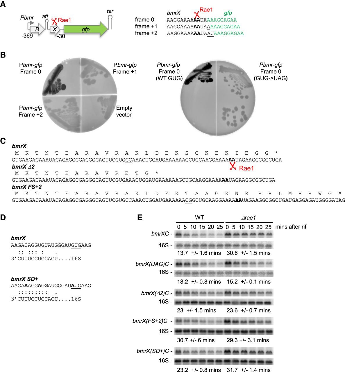
Rae1 cleaves within an unannotated cryptic ORF, bmrX, in a translation-dependent manner. (A) Structure of the pbmrBXgfp plasmid. ORFs are represented by colored boxes, the promoter by a rightward-pointing arrow, the attenuator (att) and transcription terminator (ter) by hairpin structures, and the Rae1 site by a scissors symbol. Coordinates are given relative to the AUG start codon of bmrC. The sequence of the junction of the gfp gene with the bmrBC intergenic region in the three frames is shown. The gfp sequence is indicated in green. Rae1 cleaves between the two A-residues indicated in bold. (B) Fluorescence of WT strains expressing GFP from the fusion constructs pbmrBXgfp in the three different frames (0, +1, or +2) and of the WT containing an empty vector (left panel). Fluorescence of WT strains expressing GFP in frame 0 containing either the GUG start codon or the UAG mutant (right panel). (C) Nucleotide and amino acid sequence of WT and mutant derivatives of bmrX. The Rae1 cleavage site is indicated in bold. The bmrX(Δ2) mutant contains a deletion of two C-residues (underlined in the WT bmrX sequence), creating a premature stop codon. In the bmrX(UAG) mutant, the GUG translation start codon was replaced by UAG. In the bmrX(FS + 2) mutant, 2 nt (underlined) were added to induce a +2 frameshift. (D) Ribosome binding sequence of the WT bmrX and the bmrX(SD+) mutant. The potential base-pairing with the 3′ end of 16S rRNA is indicated. The modified nucleotides in bmrX(SD+) mutant are in bold and the start codon is underlined. (E) Northern blot analysis of WT and Δrae1 strains containing WT or mutant derivatives of Pspac-bmrBXC at times after rifampicin addition, probed with oligo CC2344 (bmrC) and then with a probe complementary to 16S rRNA (CC058) as a loading control. Quantification of the blots presented is given in Supplemental Figure 4. The calculated half-lives are the average of at least three independent experiments.




