


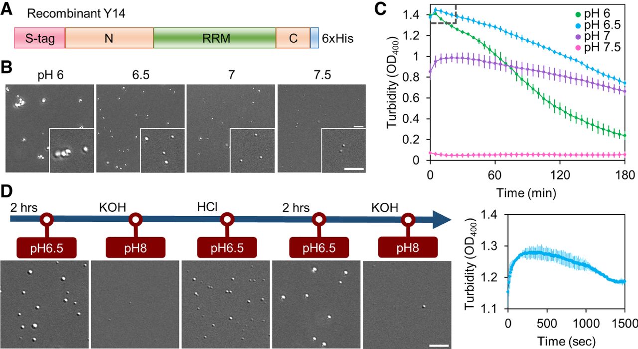
Y14 forms phase-separated droplets in a pH-dependent manner. (A) Recombinant Y14 containing the S peptide (S-tag) and hexahistidine (6 × His) tag. All recombinant Y14 variants contained these two tags. (B) In vitro LLPS of 20 µM recombinant Y14 was performed in buffers containing 50 mM KCl at different pH values for 2 h. DIC images show Y14 droplets (400 × 400 pixels; 1 pixel = 0.114942 µm); inset shows magnified images (100 × 100 pixels) of a selected area. Bar, 5 µm. The experiment was repeated more than five times (N > 5). (C) Turbidity assay showing the kinetics of LLPS of Y14 (20 µM) as in B. Turbidity at 400 nm (OD400) was measured. Optical density was recorded at 5-min intervals for 3 h (upper). Dashed square: recording was additionally taken at 10-sec intervals for the first 25 min (lower). Mean and standard deviation were calculated for two to four replicates. (D) Y14 (20 µM) was initially incubated in a pH 6.5 buffer. The pH of the buffer was subsequently adjusted by sequential addition of KOH, HCl, and KOH again, as indicated. DIC images were acquired immediately or after a 2-h incubation, as indicated. Bar, 5 µm. N = 2.




