


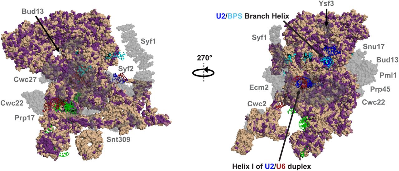
FIGURE 8.
Surface representation of the Cm Bact complex homology modeled against the Sc structure (PDB 5GM6). All protein sequences were annotated by conservation with their Cm homologs. Identical and similar amino acids are colored purple while dissimilar amino acids are colored wheat. Proteins absent in Cm are shown in transparent gray. The RNA is colored as follows: U6 (dark red); U5 (green); U2 (blue); and pre-mRNA (cyan).




