


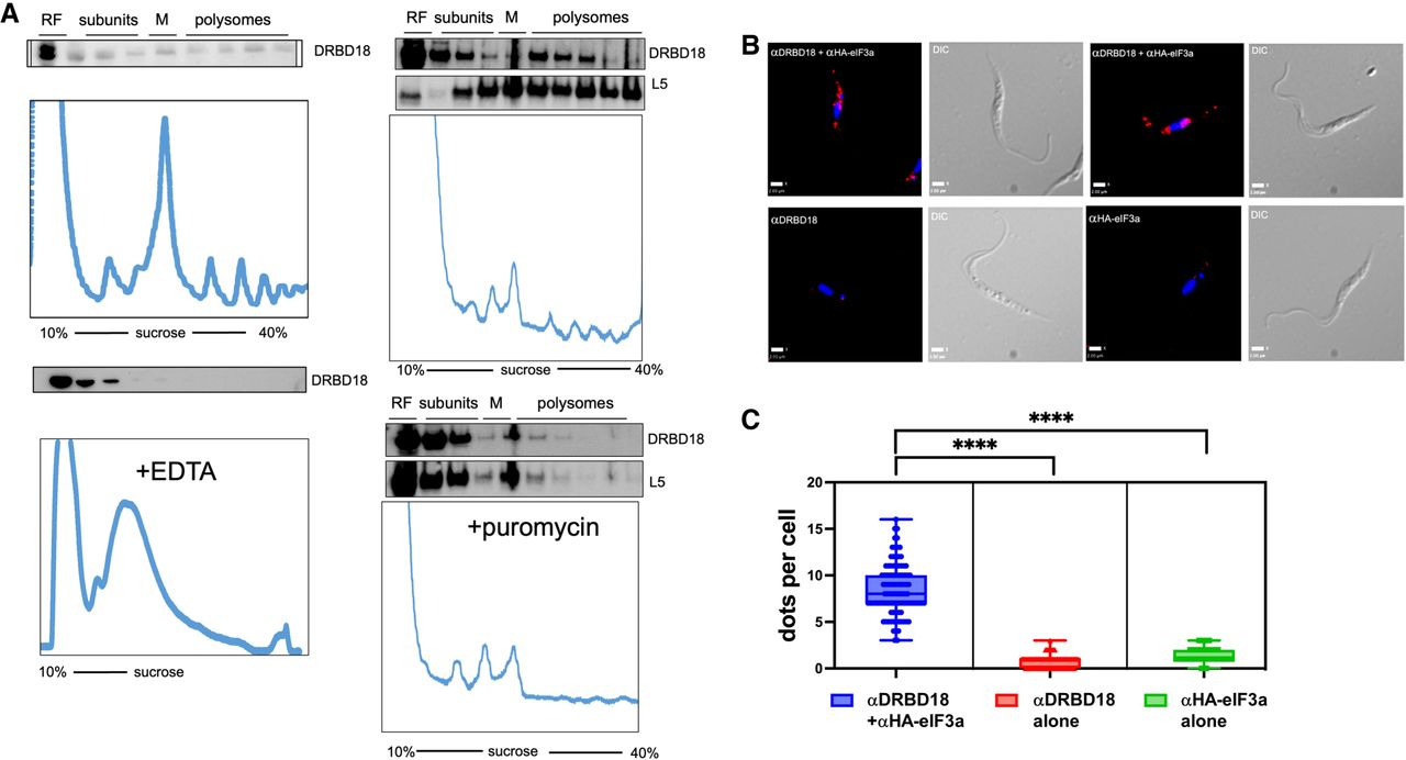
DRBD18 associates with polysomes and translation initiation factor eIF3a. (A) Anti-DRBD18 western blot detection of DRBD18 in high-density, polysomal fractions from cycloheximide-treated lysates separated on 10%–40% sucrose gradients (top). EDTA-mediated (bottom left) or puromycin-mediated (bottom right) dissociation of ribosomes leads to loss of DRBD18 association with high-density fractions. (L5) ribosomal protein L5, (RF) ribosome free, (M) monosomes. (B) Proximity ligation assay (PLA) in cells with carboxy-terminally HA-tagged eIF3a using antibodies against HA and DRBD18. Two examples are shown in the top row with corresponding DIC images. Red dots indicate signal from the amplification of individual interactions. Negative controls in the bottom row are individual antibodies alone; anti-DRBD18 (left), anti-HA (right), with corresponding DIC images. Bar, 2 μm. (C) Quantification of dots per cell (100 cells per condition); both antibodies, blue; anti-DRBD18 alone, red; anti-HA alone, green. Students t-test, (****) P < 0.0001.




