


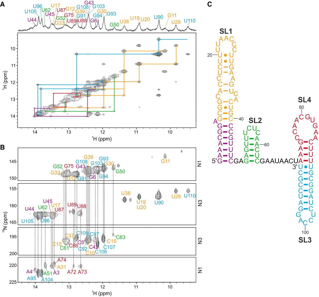
FIGURE 5.
NMR characterization of 110 nt OxyS RNA. The color codes follow the colors used in the studies of individual stem–loops. (A) Imino–imino region of JR-NOESY spectrum (τm = 200 msec) with traced imino–imino contacts, acquired at 5°C. (B) 1H–15N HNN-COSY spectrum acquired at 5°C. Resonances of donor and acceptor nitrogens involved in hydrogen bonds, specifically G(N1)-C(N3) and U(N3)-A(N1), are connected with dashed gray lines. (C) NMR-derived secondary structure of OxyS. Spectra were acquired at 1 mM RNA concentrations, 15 mM Na-phosphate buffer, 30 mM NaCl, and pH 6 in 90% H2O and 10% 2H2O on a 600 MHz NMR spectrometer.




