


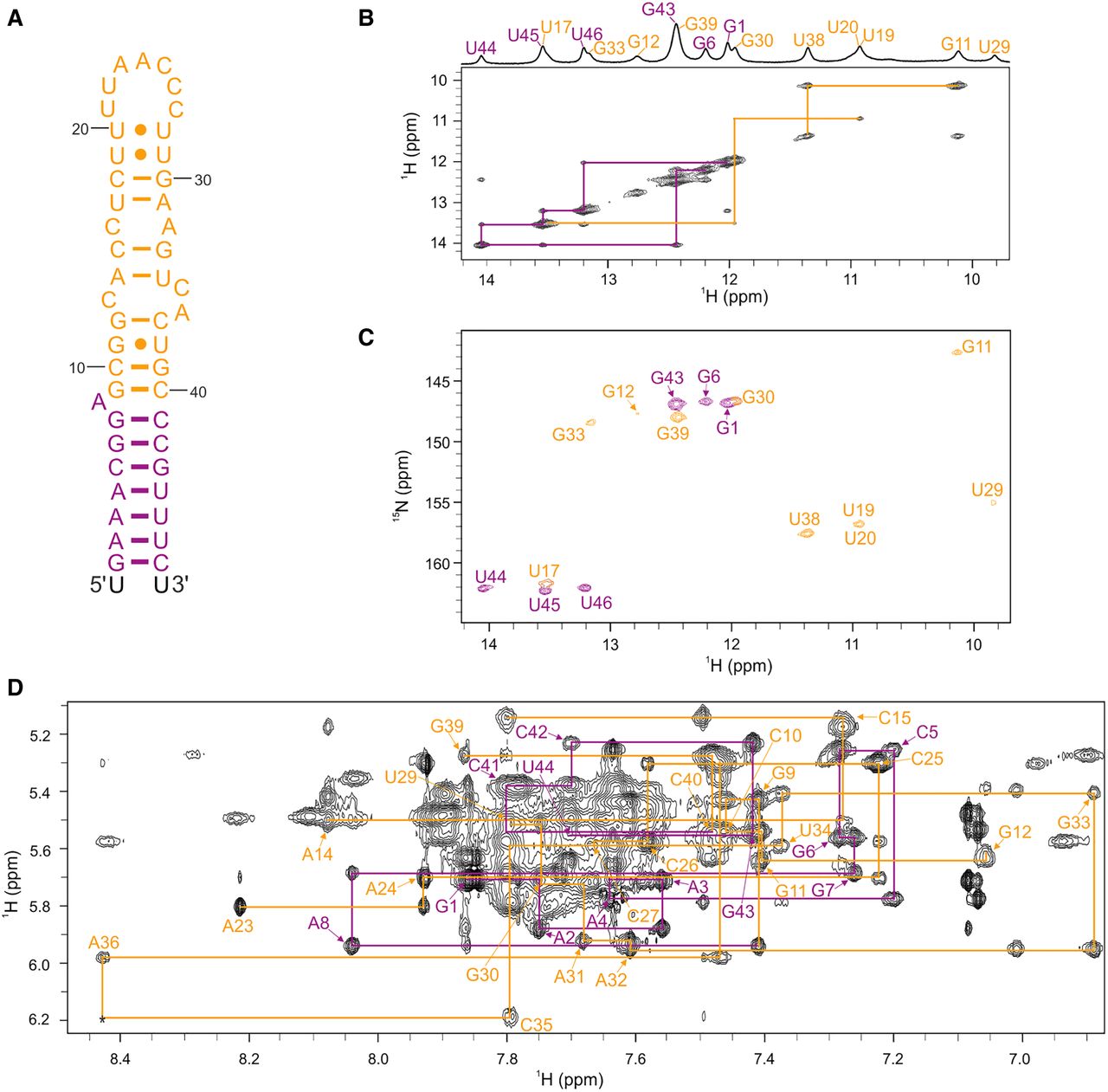
FIGURE 4.
NMR structural characterization of SL1. (A) Secondary structure of SL1. Truncated construct SL1Δ17 is colored in orange and the remaining nucleotides are in purple. Overhangs at 5′- and 3′-end are in black. (B) Imino–imino region of JR-NOESY spectrum (τm = 200 msec) of SL1, acquired at 5°C. Imino–imino contacts are denoted with purple and orange lines. (C) 1H–15N SF-HMQC spectrum of SL1, acquired at 5°C. (D) Aromatic–anomeric region of NOESY spectrum (τm = 250 msec) of SL1 with traced sequential connectivities, acquired at 25°C. Spectra were acquired at 1 mM RNA oligonucleotide, 15 mM Na-phosphate buffer, 30 mM NaCl, and pH 6 in 90% H2O and 10% 2H2O on 600 and 800 MHz NMR spectrometers.




