


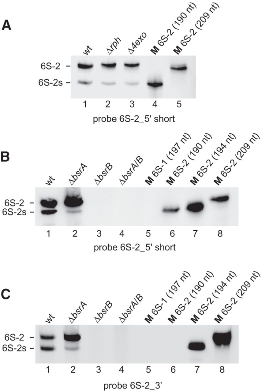
Northern blot analysis of a 6S-2 RNA degradation intermediate termed 6S-2s using the probes as indicated below each blot. (A) The presence of the 6S-2s signal is detectable (lane 1) with probe 6S-2_5′ short (see Fig. 9), indicating that 6S-2s RNA is shortened at the 3′-end; 6S-2s RNA is also detectable in the Δrph and Δ4exo strains (lanes 2,3), indicating that it is generated by endonucleolytic cleavage rather than 3′-exonucleolytic degradation of 6S-2 RNA. (B) Detection of 6S-2s RNA by probe 6S-2_5′ short in a B. subtilis PY79 6S-1 RNA deletion strain (ΔbsrA; lane 2), but not in a 6S-2 RNA deletion strain (ΔbsrB; lane 3) or a 6S-1/2 double deletion strain (ΔbsrA/B; lane 4). (C) As in panel B but using probe 6S-2_3′ (Table 1; Fig. 9). This probe detects the 194 nt 6S-2 marker RNA (lane 7) that has its 3′-end at position 193 (Fig. 9), but not the 190 nt marker RNA with the 3′-end at position 189 (lane 6). For the sequence of 6S-1 and 6S-2 marker RNAs, see the Supplemental Material.




