


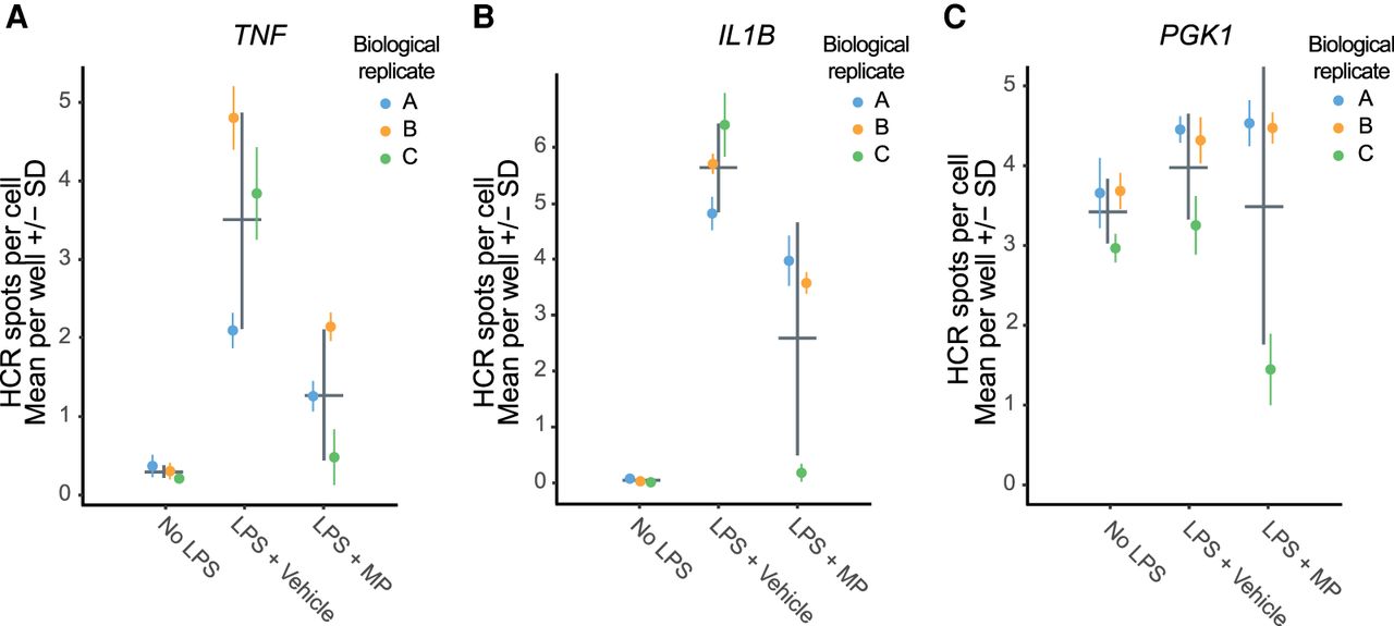
Assessment of technical and biological variation in hcHCR data. Human primary monocytes were obtained from three unrelated healthy donors (biological replicates) and hcHCR was performed with TNF (A), IL1B (B), or PGK1 (C) as the target mRNAs in 12 wells per donor (technical replicates). Measurements were performed at baseline (no LPS), after stimulation with 1 ng/mL LPS for 30 min followed by vehicle treatment (0.1% EtOH) for 2 h (LPS + Vehicle), or after stimulation with 1 ng/mL LPS for 30 min followed by methylprednisolone treatment (200 µg/dL) for 2-h (LPS + MP). Each dot represents one biological replicate. Colored error bars display the mean ± SD of the 12 technical replicates measured in each biological replicate. Gray error bars display the mean ± SD of the biological replicates.




