


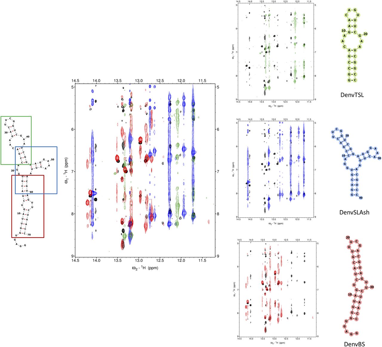
FIGURE 3.
Overlay of the imino region of NOESY spectra for DenvSLATL (black) and the three smaller constructs which were prepared to facilitate structure determination. Black color corresponds to DenvSLATL, red to DenvBS, the bottom helix; green corresponds to DenvTSL, the top stem–loop, and blue corresponds to DenvSLAsh, the three-way junction. 1H NOESY spectra were recorded in 10 mM potassium phosphate buffer (pH 6.5, 90% H2O/10% D2O) at 25°C. Despite small differences, a high similarity of chemical shifts and NOE patterns were observed, allowing the transfer of peak assignments from each segment to the SLA model oligonucleotide (Supplemental Fig. S3).




