


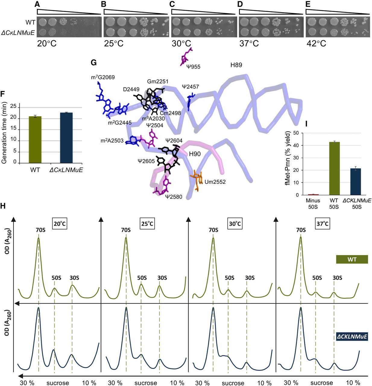
Assays of growth rate, ribosome sucrose gradient profile and peptidyl transferase with ΔCKLNMuE. (A–E) Representative spot growth assays on chloramphenicol agar plates (see Materials and Methods). Both strains carried a control plasmid (empty vector carrying CmR). (F) Generation times of liquid cultures at 37°C (standard errors of eight biological replicates). (G) 3D proximity of all modified nucleotides of domain V (Borovinskaya et al. 2007) colored according to their respective modification enzymes in Figure 1A. ΔCKLNMuE lacks the modifications on the 8 nts colored blue and purple (and also lacks m2A37 modifications in six tRNAs, which are not shown). The entire backbone of the critical region is given (blue), as is the backbone that base pairs with it in H90 (purple; see Fig. 1A). (H) Ribosome sucrose gradients of cells grown at the temperatures indicated, with representatives of two biological replicates shown. (I) Representative plots of peptidyl transferase puromycin “fragment” reactions in vitro (standard errors of three technological replicates).




