


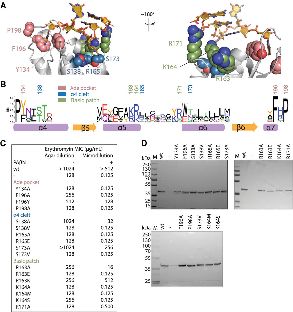
Erythromycin resistance phenotypes of site-directed mutants of the adenosine pocket, the α4 cleft, and the basic ridge. (A) A detailed view of the theoretical model of ErmE bound to Helix 73 of rRNA is shown. The model predicts residues involved in noncovalent interactions driving RNA recognition. 23S rRNA is shown in gold and selected amino acids are colored based on which region of ErmE they reside in: the adenosine pocket (pink), the α4-cleft (blue), and the basic patch (green). (B) One representative from each class of Erm proteins was used to model the sequence conservation at the three sites of interest. (C) Minimal inhibitory concentrations (MIC) for erythromycin were measured for E. coli cells expressing site-directed mutants of ErmE. Experiments were conducted as agar dilution without the antibiotic adjuvant phenyl-arginyl-beta-napthylamide (PAβΝ) or as liquid culture microdilution experiments in the presence of PAβΝ. (D) When cells displayed an erythromycin-sensitive phenotype, western blotting was used to verify that ErmE site-directed mutants were expressed and stable.




