


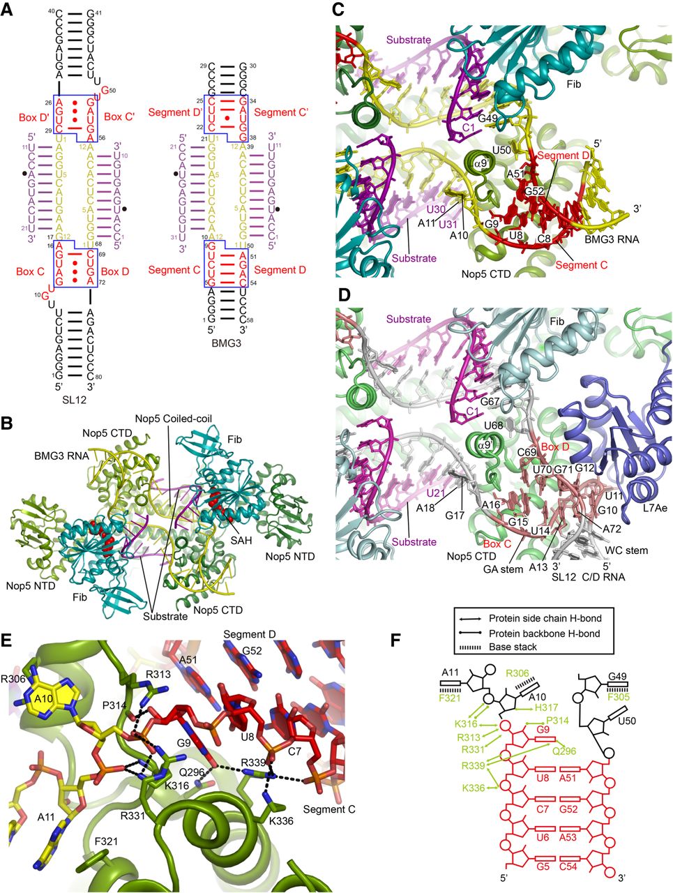
Crystal structure of substrate-bound BMG3 RNP. (A) Secondary structure diagrams of SL12 and BMG3 RNAs. C/D motifs in SL12 and segments C and D in BMG3 are colored in red, spacers in yellow and substrates in purple. Modification sites are marked by circles. The structurally equivalent Nop5-binding regions are encircled with blue boxes. Nucleotides in guide RNAs and substrates are separately numbered. Spacer sequences are additionally labeled by yellow numbers to indicate the distance to box D/D′ or segment D/D′. (B) Ribbon representation of structure of BMG3 in complex with Nop5, fibrillarin (Fib) and substrate. Two Nop5 molecules are colored in forest green and split pea, Fib in teal, guide RNA in yellow and substrates in purple. SAH bound in the active site of Fib is shown as red spheres. (C,D) RNA–protein interfaces in the BMG3 RNP (C) and SL12 RNP (PDB code: 3PLA) (D) structures. (E) Detailed interactions between BMG3 RNA and Nop5 at segments C/D. Hydrogen bonds are denoted by dashed lines. (F) Schematic of interactions between BMG3 RNA and Nop5.




