


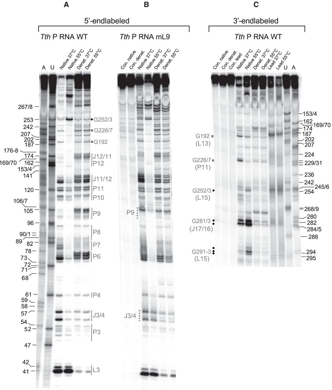
RNase T1 probing of 5′-endlabeled T. thermophilus wild-type (WT) (A) and mL9 mutant P RNA (B), and 3′-endlabeled WT P RNA (C). Lanes A and U: AMPαS- or UMPαS-substituted T. thermophilus WT P RNA used to generate A- and U-specific ladders by iodine hydrolysis; lanes “Native 37°C” and “Native 55°C”: P RNA preincubated for 10 min at 37°C or 55°C in the nondenaturing buffer B before addition of RNase T1 and limited hydrolysis for 10 min at 37°C; lanes “Denat. 37°C” and “Denat. 55°C”: P RNA preincubated for 10 min at 37°C or 55°C in the denaturing buffer A before addition of RNase T1 and limited hydrolysis for 10 min at 37°C; lanes “Con. native” and “Con. denat.”: P RNA preincubated for 10 min at 55°C in buffer B or buffer A, respectively, followed by incubation 10 min at 37°C without RNase T1. Cleavage fragments generated by iodine hydrolysis (lanes A and U) are assigned at the left (A) and right (C) margins according to the T. thermophilus P RNA numbering system shown in Figure 1C; note that shorter fragments of A and U ladders in panel C show double bands, which we attribute to end heterogeneity of the 3′-endlabeled P RNA. In A and C, regions of T. thermophilus P RNA that showed reduced or enhanced RNase T1 accessibility after preincubation at 55°C versus 37°C are marked by gray lettering as well as open and filled circles, respectively, or gray vertical lines; in C, structural elements are given in parentheses on the left, below the indicated G residues; J17/16: junction between P17 and P16. Differences in the RNase T1 protection pattern between T. thermophilus wt and mL9 P RNA are marked by vertical gray-dotted lines in B. The following regions became protected from RNase T1 cleavage upon preincubation of 5′-endlabeled Tth P RNA at 55°C versus 37°C (A): P11 (3′-strand, G226/227), L13 (G192), J11/12 (G127/129/132/134), P10 (114/5, weakly), P9 (G101–104), P6–8 (G74–76; G79–81; G87/88/93), P4 (G60), J3/4 (G55/56), and the 3′-side of P3 (G49/50/51/53). 3′-endlabeled Tth P RNA (C) provided partially overlapping information, such as protection at G226/7 or signal enhancement (filled dots) at G252/3 upon preincubation at 55°C; signal intensities were also increased for cleavages in the 3′-part of L15 (G291–3) and in J17/16 (G281/3).




