


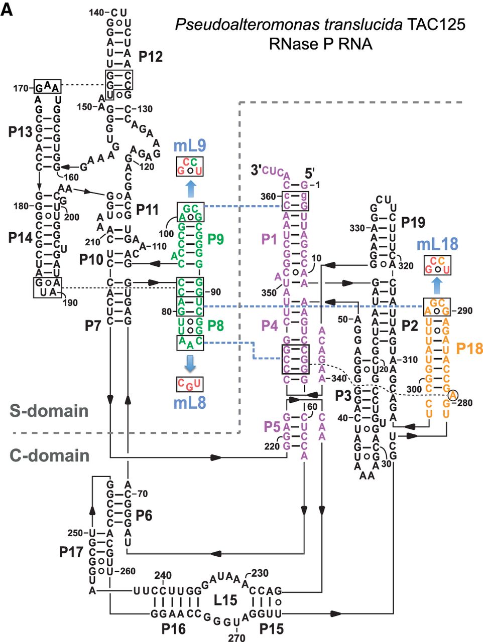
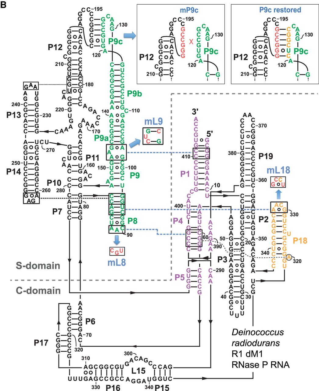
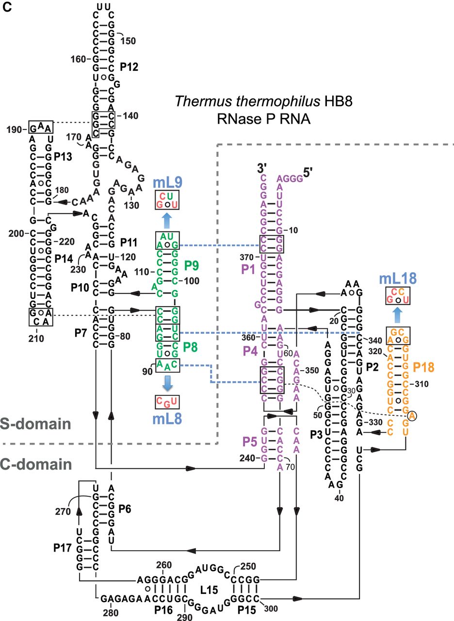
Secondary structure presentation of (A) P. translucida (strain TAC125), (B) D. radiodurans (strain R1 dM1) and (C) T. thermophilus (strain HB8) P RNAs according to Massire et al. (1998), with the three interdomain tetraloop-helix contacts L18–P8, L8–P4, and L9–P1 indicated by dashed blue lines and the involved structural elements in color. Nucleotide exchanges in L8, L9, and L18 (mL8, mL9, and mL18) that disrupt these tertiary interactions are highlighted in red. In the Dra P RNA, the mP9c and “P9c_restored” mutations are illustrated in the boxes. In the P. translucida P RNA (A), the second bp in P1 is shown in small letters to indicate that the bp was mutated from A:U to G:C (for details, see text). Numbering in C is according to Schlegl et al. (1994); numberings in A and B are based on the rnpB genes annotated in the P. translucida strain TAC125 chromosome I (GenBank CR954246.1) and the D. radiodurans strain R1 dM1 chromosome I (GenBank CP031500.1), respectively.




