


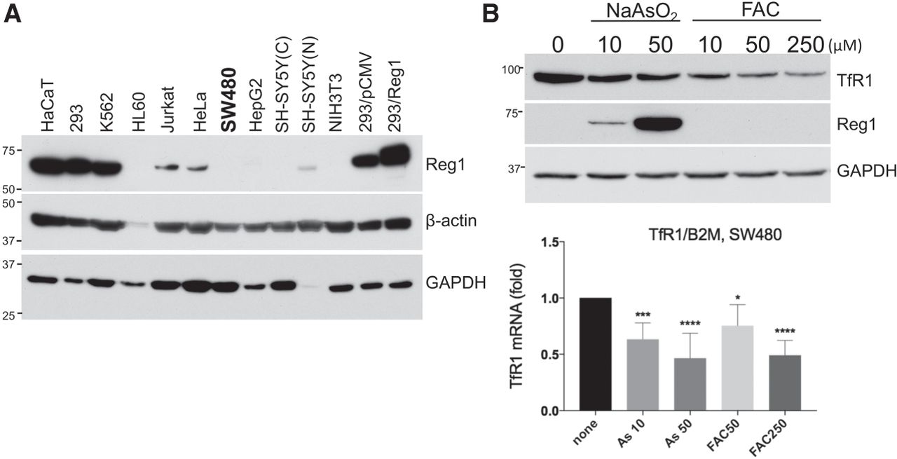
Reg1 expression in various cell lines and the effects of Reg1 on endogenous TfR1 expression. (A) Thirty micrograms of whole cell lysates isolated from nine cell lines and cytoplasmic (C) and nuclear (N) fractions from SH-SY5Y cells were loaded on SDS-PAGE and subjected to western blotting with anti-ZC3H12A (Reg1) antibody (GeneTex, GTX110807), anti-β-actin antibody (SIGMA, A5441), and anti-GAPDH (Chemicon, MAB374). (B) SW480 cells were treated with 10, 50 µM NaAsO2, or 10, 50, 250 µM FAC (ferric ammonium citrate) for 18 h and harvested for preparation of whole cell lysates and total RNA. (Top) TfR1 protein expression was detected by western blotting with anti-TfR1 antibody (Abcam ab84036), followed by Reg1 and GAPDH antibodies. (Bottom) Total RNA was subjected to cDNA synthesis and qPCR according to Miyazawa et al. (2018). TfR1 Cq was normalized with B2M Cq (β2-microglobulin), and eight qPCR results from three experiments were statistically analyzed by one-way ANOVA with Prizm 7. P-values compared to none; (*) P = 0.0124, (***) P = 0.0002, (****) P = 0.0001.




