


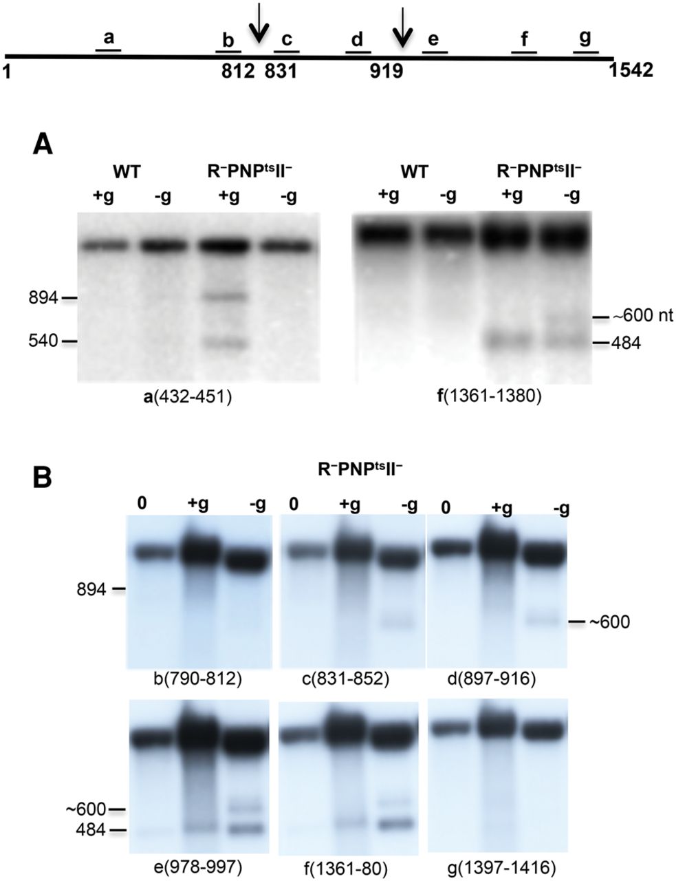
Northern blot analysis of 16S rRNA from WT and exoribonuclease-deficient cells. Total cellular RNA was extracted from cells first grown in M9/glucose at 31°C to early exponential phase (0 time) and then either starved for 4 h (−glucose) or grown for 4 h (+glucose) at 42°C. The RNA was resolved by 1% agarose gel electrophoresis, then transferred to a nylon membrane and hybridized to specific oligonucleotide probes against 16S rRNA, as indicated. (A) Oligonucleotide probes a and f were hybridized to the membrane. The probes are complementary to16S rRNA residues as indicated below each picture. (B) Probes b, c, d, e, f, and g were hybridized to the membrane. The diagram at the top (not to scale) shows the location of the probes used. Arrows represent the sites of endonuclease cleavages in 16S rRNA.




