


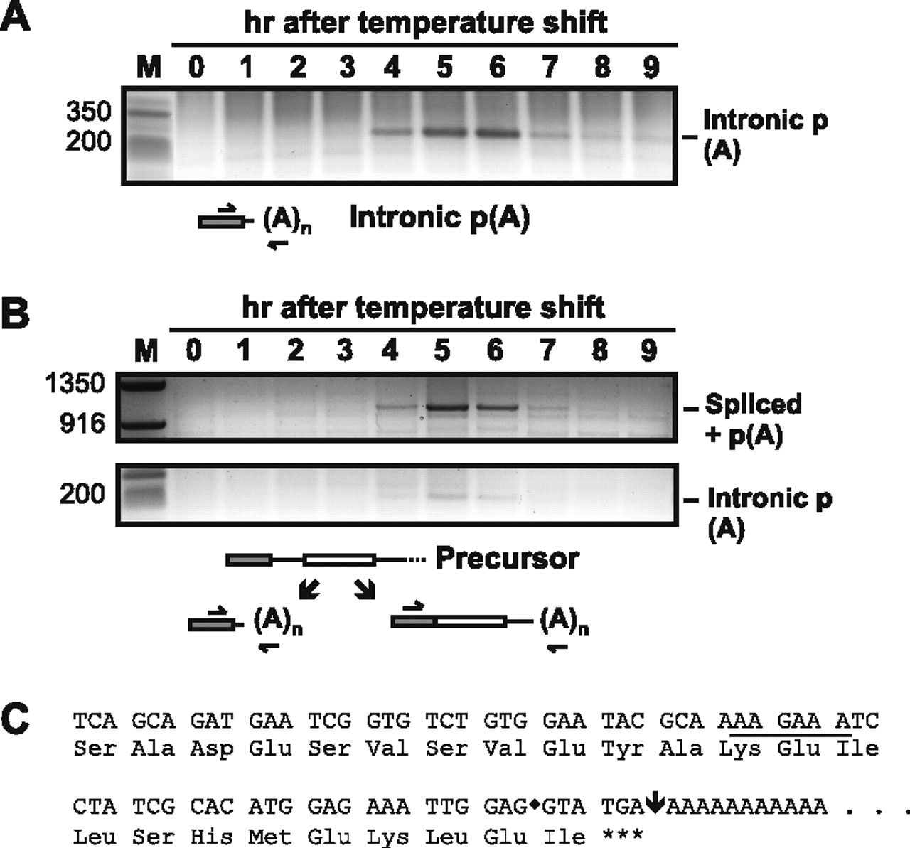
(A) Discovery of a polyadenylation site within the rem1 intron. To optimize conditions for detecting a truncated rem1 mRNA, PCR amplification was performed with a shortened extension time for each cycle. The positions of primers are diagrammed beneath the gel image. (B) Simultaneous detection of intronic and downstream polyadenylation. Once we knew its size, we were able to detect the product derived from polyadenylation within the intron using standard RT-PCR and gel conditions (see Materials and Methods); the same 5′ primer also detects the (much larger) spliced product that is polyadenylated at the downstream site as diagrammed at the bottom. Two slices from the top third and bottom quarter of the same gel are shown. (C) Sequence analysis to identify the position of intronic polyadenylation. The bands marked intronic p(A) in panels A and B were eluted from the gels, and the sequence extending into the poly(A) tail was determined. Both gave the sequence shown, in which the exon/intron boundary is marked with a diamond. As indicated by the conceptual translation, the intron lies between codons such that the unspliced RNA polyadenylated within the intron encodes a single isoleucine beyond the peptide translated from exon 1 before a termination codon is encountered immediately preceding the poly(A) tail.




