


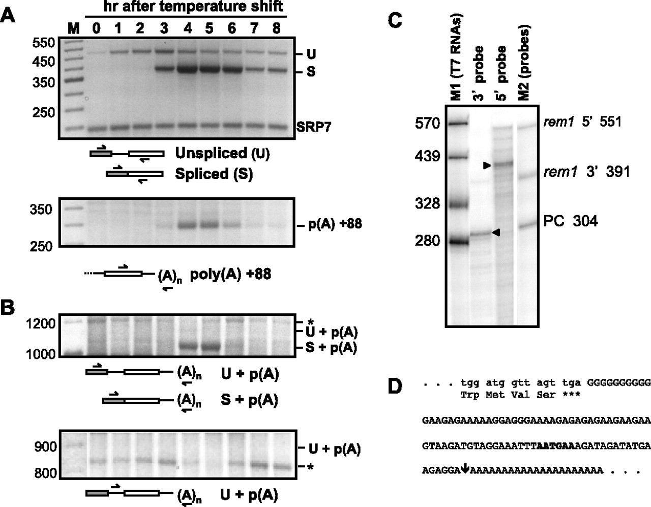
(A) Splicing (top) and polyadenylation (bottom) of rem1 transcripts peak concurrently during meiosis. RNA processing was assayed by RT-PCR as diagrammed beneath each gel image. The identities of the processed and unprocessed products, which were verified by eluting the bands and subjecting them to sequence analysis, are indicated alongside the gel image. SRP7, the small stable cytoplasmic RNA component of the signal recognition particle (Brennwald et al. 1988), was used as an internal loading control for splicing assays (Webb and Wise 2004). The 3′ primer used to assay polyadenylation (bottom) was oligo(dT) (McPheeters et al. 2009). (B) Polyadenylated transcripts that retain the intron are below the level of detection. Splicing and 3′ processing were assayed concurrently using a 5′ primer complementary to exon 1 (top) or the intron (bottom). Asterisks mark bands that did not yield readable sequence with rem1-specific primers. (C) Mapping the 5′ and 3′ termini of rem1 RNA using an RNAse protection assay. Total RNA prepared from mid-meiotic cells was incubated with uniformly labeled riboprobes extending beyond either end of the transcript. The sizes of the major digestion products, indicated by arrowheads, were determined graphically based on mobility relative to the two sets of markers. Faint bands comigrating with the undigested 5′ and 3′probes (right lane) remain in each lane. (PC) a positive control probe complementary to a mouse mRNA supplied by the manufacturer; removal of the lane displaying this assay is indicated by a white line. (D) Sequence of the region extending from the final four codons of the open reading frame through the polyadenylation site. The location of the poly(A) tail addition was determined by sequence analysis of RT-PCR products eluted from multiple gels including the one shown in panel A, with reproducible results. The site of cleavage, marked with a downward arrow, may be specified by the sequence highlighted in bold, which is a putative positioning element (see text for details).




