


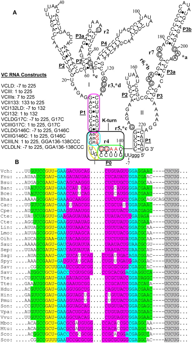
(A) Secondary structure of the full-length VC glycine riboswitch. r2–r7; *a–*b indicate the representative glycine-induced perturbation regions in the in-line probing assay and the hydroxyl radical footprinting assay of VCIII, respectively; the corresponding nucleotides are circled. (I, II) Aptamer I and aptamer II, respectively. Green, yellow, cyan, purple, and gray regions indicate C stem (P0 helix), internal loop, NC stem, P1 helix of aptamer I, and P1 helix of aptamer II, respectively. The kink-turn motif is shown in a black box. Red nucleotides indicate previously defined linker sequence. (B) Alignment of the kink-turn motifs formed between leader and linker regions in 31 of 55 tandem glycine riboswitch sequences reported by Breaker and coworkers (Mandal et al. 2004). Abbreviations of the names of the organisms are taken from the same article.




