


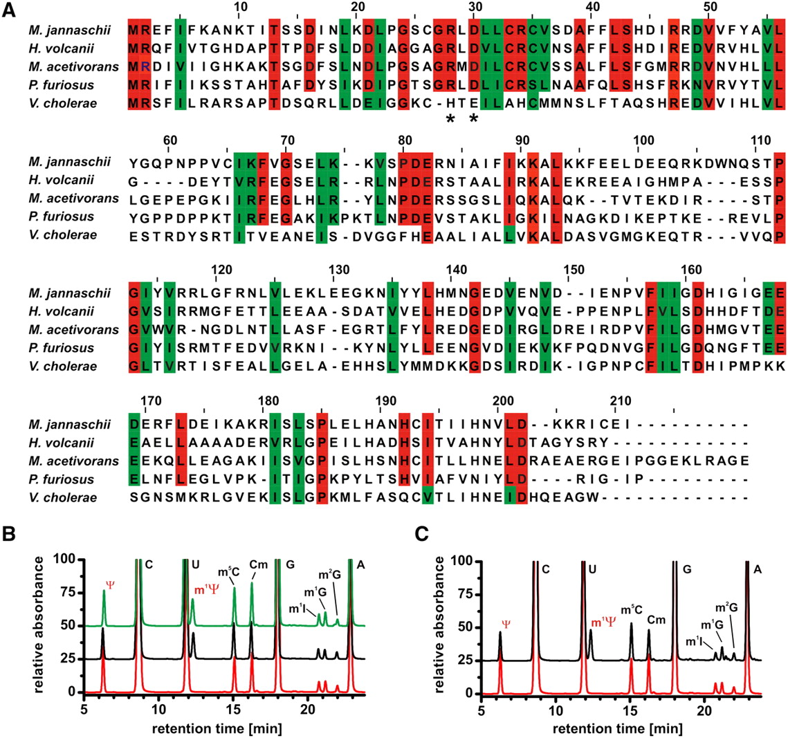
Hvo_1989 activity in vivo. (A) Hvo_1989 and Mja_1640 are highly homologous proteins, as seen from the sequence alignment of members of the DUF 358 family. Identical amino acids of the archaeal members of this family are colored in red, similar amino acids are shown in green. The arginine and the aspartate important for pseudouridine recognition and orientation are indicated by a star. Only in the bacterial member of this family these amino acids are a histidine and a glutamate, respectively. (B) Nucleoside analysis of unfractionated tRNAs isolated from wild-type (black) and the in-frame deletion mutant cells (red) and mutant cells complemented with the pSD1/M2-18 plasmid (green). For the tRNAs from wild-type and complemented strains the N1-methyl pseudouridine signal (indicated in red) is clearly present, while the signal is absent in the case of the in-frame deletion mutant. The putative assignments for the signals corresponding to other modified nucleosides are also indicated (Edmonds et al. 1991). (C) Nucleoside analysis of tRNAs isolated from in-frame deletion mutant cells (red) and the same tRNAs after incubation with SAM and Mja_1640 (black). The N1-methyl pseudouridine signal only appears after incubation with Mja_1640.




