


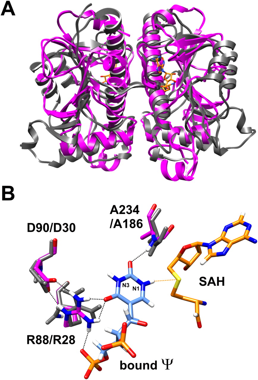
Structural similarity between Nep1 and Mja_1640. (A) Overlay of the structures of the MjNep1 (pdb 3bbe) and Mja_1640 (pdb 3aia) dimers revealing the similar overall fold typical for SPOUT-class RNA methyltransferases. (B) Model of pseudouridine bound to the active center of Nep1 derived from the structure of yeast Nep1 bound to S-adenosylhomocysteine and an inhibitor RNA containing uridine instead of pseudouridine at the reactive position (pdb 3oin). The amino acids R88, D90, and A234, which form hydrogen bonds to the pseudouridine carbonyl groups and orient the N1 nitrogen toward the sulfur atom of the cofactor, are colored by atom types. The equivalent amino acids in Mja_1640 are shown in gray and numbered according to the Mja_1640 numbering scheme.




