


Activation of PKR by RNA misfolding: HDV ribozyme dimers activate PKR
- Department of Chemistry, Center for RNA Molecular Biology, The Pennsylvania State University, University Park, Pennsylvania 16802, USA
Abstract
Protein Kinase R (PKR), the double-stranded RNA (dsRNA)-activated protein kinase, plays important roles in innate immunity. Previous studies have shown that PKR is activated by long stretches of dsRNA, RNA pseudoknots, and certain single-stranded RNAs; however, regulation of PKR by RNAs with globular tertiary structure has not been reported. In this study, the HDV ribozyme is used as a model of a mostly globular RNA. In addition to a catalytic core, the ribozyme contains a peripheral 13-bp pairing region (P4), which, upon shortening, affects neither the catalytic activity of the ribozyme nor its ability to crystallize. We report that the HDV ribozyme sequence alone can activate PKR. To elucidate the RNA structural basis for this, we prepared a number of HDV variants, including those with shortened or lengthened P4 pairing regions, with the anticipation that lengthening the P4 extension would yield a more potent activator since it would offer more base pairs of dsRNA. Surprisingly, the variant with a shortened P4 was the most potent activator. Through native gel mobility and enzymatic structure mapping experiments we implicate misfolded HDV ribozyme dimers as the PKR-activating species, and show that the shortened P4 leads to enhanced occupancy of the RNA dimer. These observations have implications for how RNA misfolding relates to innate immune response and human disease.
Keywords
INTRODUCTION
Hepatitis delta virus (HDV) is a satellite virus of hepatitis B virus (HBV), where coinfection by HDV leads to a more virulent form of the infection (Lai 1995; Lazinski and Taylor 1995; Karayiannis 1998). The 1.7-kb circular, single-stranded RNA genome of HDV is responsible for making genomic and antigenomic strands of RNA. Replication of the HDV genome is assisted by self-cleavage of a semiglobular (Ferre-D'Amare et al. 1998), ∼84-nt ribozyme located in both genomic and antigenomic RNA strands, which serves to linearize concatamers. Sequence downstream from the ribozyme, referred to as the “attenuator” sequence, sequesters native ribozyme pairings and forms a long rod-like structure (Fig. 1; Wang et al. 1986; Lazinski and Taylor 1993, 1995).
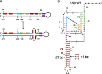
Secondary structures of HDV genome segment and HDV ribozyme. (A) Portion of the HDV genome illustrating rod-like (top) and alternative cruciform (bottom) structures (Branch and Robertson 1991; Diegelman-Parente and Bevilacqua 2002). Color-coding represents two strands that either base pair in the ribozyme (colors match those in B) or in the upstream P(−1) region between −54 and −1 (Chadalavada et al. 2000). (B) Secondary structure for 1/99 WT and alterations in the 1/99 P4 variants, Δ3 bp and +3 bp. Variant 1/99 Δ3 bp was prepared by removing the following six nucleotides: G50, A51, G52, C64, U65, and C66 (all six nucleotides boxed), while 1/99 + 3 bp was prepared by adding the six nucleotides at the position shown.
Two previous studies characterized PKR activation by a 482-nt segment of the HDV genomic RNA (annotated as −207/275, where 1 is the first nucleotide of the ribozyme) (Robertson et al. 1996; Circle et al. 1997). This region of RNA includes the ∼84-nt ribozyme and forms its rod-like structure due to native ribozyme base pairs being sequestered by attenuator sequence (Fig. 1A, top structure). Within this rod-like structure, there are no stretches of dsRNA greater than 20-bp. In addition to reporting activation of PKR by this long segment of HDV, the investigators mapped the PKR-binding site to the ribozyme-containing portion of the rod-like structure. This particular site has also been reported to form a cruciform structure (Fig. 1A, bottom structure; Branch and Robertson 1991; Diegelman-Parente and Bevilacqua 2002).
Prior studies have shown that PKR is activated by long dsRNAs, typically >33 bp in length (Manche et al. 1992; Zheng and Bevilacqua 2004). In addition, PKR is known to be activated by highly structured RNA domains of hepatitis C virus (HCV) (Shimoike et al. 2009; Toroney et al. 2010) and a pseudoknotted human IFN-γ-mRNA (Ben-Asouli et al. 2002; Cohen-Chalamish et al. 2009). Unlike the above activators, the HDV ribozyme is semiglobular, although it contains a protruding 13-bp pairing region (P4). Globular RNA activators of PKR with the characteristics of HDV have not been previously examined.
In this report, we focus on the regulation of PKR by the 1/99 HDV ribozyme sequence—a much smaller fragment of the previously reported PKR-regulating construct, and one that is missing the attenuator—and investigate the role of the P4 region in activation. Variants of 1/99 were prepared with shortened or lengthened P4 regions, with expectation that lengthening P4 would lead to increased PKR activation since it would offer more base pairs of dsRNA. Surprisingly, “shortening” P4 increased PKR activation. Herein, we report that ribozyme dimers, with an extended kissing loop, are the activating species and that shortening P4 enhances the extent of dimer formation and PKR activation. We discuss possible biological implications of this activity.
RESULTS AND DISCUSSION
Activation of PKR by monomer/dimer mixtures of 1/99 HDV variants
Our lab has been interested in identifying and characterizing RNA motifs that regulate PKR activity (Zheng and Bevilacqua 2004; Nallagatla et al. 2007; Heinicke et al. 2009, 2011; Toroney et al. 2010); we thus chose to examine activation of PKR by the semiglobular HDV ribozyme. Various lengths of the HDV genome, including wild-type uncleaved and cleaved, and the catalytically unreactive C75U forms of –54/271, –54/186, –54/172, –54/140, and –54/99 were initially tested for PKR activation. All RNAs activated PKR to some extent, with optimal RNA concentrations of 0.5 μM for longer RNAs and 5 μM for shorter RNAs. There was no clear trend in the ability of WT versus C75U to activate PKR (Heinicke 2010), suggesting that the activating species is not the monomeric RNA with catalytic tertiary structure. We chose to focus this study on the 1/99 WT construct, which was the minimal RNA motif able to activate PKR.
Biochemical and structural analyses indicate that this region is mostly globular, but with a long protruding pairing termed P4 (Fig. 1; Ferre-D'Amare et al. 1998; Chadalavada et al. 2000). Early catalytic studies on the HDV ribozyme showed that shortening P4 does not significantly affect catalytic activity (Been and Wickham 1997) and that P4-shortened forms readily crystallize in catalytically relevant conformations (Ferre-D'Amare et al. 1998). The P4 loop sequence (L4) is largely self-complementary (ACCGU); thus, we hypothesized that two molecules of HDV may interact through L4 as kissing hairpins. This interaction would make a ∼28-bp dsRNA segment onto which two PKR molecules could bind and autophosphorylate. Such RNA lengths are near the minimal activating length of ∼33 bp (Manche et al. 1992; Zheng and Bevilacqua 2004). Thus, if this model were correct, shortening P4 would decrease PKR activation, while lengthening it would increase activation. Subsequent experiments would prove that this model is too simple.
We designed a shortened P4 variant with 3 bp removed, referred to as “1/99 Δ3 bp,” and a lengthened P4 variant that has 3 bp inserted, referred to as “1/99 + 3 bp” (Fig. 1B). (Note that we avoided weakening P4 through the introduction of bulges, as this would result in bulges in the concomitant dimers, which would very likely prevent PKR activation as recently shown [Heinicke et al. 2011].) For this study, all three 1/99 variants were prepared from self-cleaved transcripts that begin at –54 and end with 99 and were isolated on a denaturing 6% PAGE. The extent of cleavage during transcription was extensive, with –54/99 Δ3 bp cleaving up to ∼80%, and –54/99 WT and –54/99 + 3 bp cleaving to ∼90%. These extents of cleavage provided adequate cleaved ribozyme for the present study.
The three HDV variants were examined for sample heterogeneity by native gel-mobility analysis (Fig. 2A). Monomeric HDV ribozyme is known to be well-folded in 200 mM NaCl and 10 mM MgCl2 (Brown et al. 2004); however, effects of salt on HDV multimerization have not been investigated. We examined sample heterogeneity of the total sample in three salt conditions: First, RNA was denatured at 90°C for 1 min, then incubated at 55°C for 10 min, followed by incubation at room temperature for 10 min, and then treated with an equal volume of (1) TE, (2) TEK400M20, or (3) TEN400M20 (see Materials and Methods for buffer shorthand), such that the final salt for (2) and (3) was 200 mM KCl (or NaCl) and 10 mM MgCl2. As shown in Figure 2A, changing the salt did not affect electrophoretic mobility, but increasing the RNA concentration yielded more dimer for all variants, consistent with Le Châtelier's principle (Fig. 2A, cf. lanes 6–8 with 3–5, lanes 12–14 with 9–11, or lanes 18–20 with 15–17). (See Materials and Methods for confirmation of monomer and dimer species according to tRNA markers.) Notably, 1/99 Δ3 bp formed the most dimer (∼50%), while 1/99 WT and 1/99 + 3 bp formed only small amounts of dimer (∼10%). Increased dimer formation in 1/99 Δ3 bp is likely due to destabilization of P4 in the monomeric form. We note that enhanced dimerization of the shortened form of the ribozyme does not contradict the results of Been and Wickham (1997), that shortening P4 does not affect catalytic activity in that their experiments were conducted with trace amounts of radiolabeled RNA, which are unlikely to dimerize, while our experiments were conducted with 20 μM of RNA (Fig. 2A).
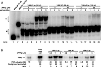
Native gel analysis of HDV dimerization and its association with PKR activation. (A) Native gel analysis of RNA. Gel is 10% native PAGE run in THEM4. 1/99 Δ3 bp, WT, and +3 bp were renatured at 2 or 20 μM concentrations with trace 5′-end labeled RNA in TE, by incubating at 90°C for 1 min, followed by room temperature for 10 min, then 55°C for 10 min, followed by incubation at room temperature for 10 min. The sample was then mixed with an equal volume of one of the following salt conditions (labeled in gel): (1) TE, (2) TEK400M20, or (3) TEN400M20; final ionic concentrations in 2 and 3 were 200 mM KCl (or NaCl) and 10 mM MgCl2. Number of nucleotides in cleaved monomer HDV is provided above the gel. The faster migrating bands are assigned as monomer (M) and the slower migrating bands as dimer (D) as described in the Materials and Methods; assignment of these bands is facilitated by structure mapping (see below) and by comparison to lanes 1 and 2, which are tRNA monomer (M) and dimer (D), with the dimer prepared as per Wittenhagen and Kelley (2002) and Roy et al. (2005). (The minor, faster-mobility band associated with M and D was not always present in native gels [e.g., absent in Figs. 3A, 4B] and may represent alternative RNA structures induced by the presence of divalent salt in THEM4 buffer, which is absent in Figs. 3A, 4B.) (B) PKR activation by HDV variants containing a mixture of the M and D species. Renaturation and ionic conditions are provided in the Materials and Methods. A no-RNA lane is provided, and phosphorylation activities are normalized to 0.01 μM 79 bp RNA. Both no-RNA and 79-bp RNA lanes contain a final concentration of 100 mM NaCl and 5 mM MgCl2. The no-RNA lane was subtracted from each lane in order to provide RNA-dependent activation values.
To test for a correlation between HDV sample multimerization and PKR activity, we performed activation assays using RNA that had been renatured in the ways that provide the various species of Figure 2A using final conditions of TEN100M5. As shown in Figure 2B, the 1/99 Δ3-bp variant, which was left as a monomer/dimer mixture in this instance, activated PKR significantly more potently than 1/99 WT or 1/99 + 3 bp (13- to 34-fold more in the presence of 1 μM RNA). These trends in activation correlate with the greater amount of dimer in the native gel (Fig. 2A), implicating dimer in PKR activation. Also, consistent with previous reports, PKR activation exhibits a bell-shaped dependence on RNA concentration for the most potent RNA activator (see Fig. 2B, 1/99 Δ3 bp). The bell shape arises because high concentrations of dsRNA activators titrate PKR into monomers, thus preventing PKR dimerization and activation (Lemaire et al. 2008).
Activation of PKR by purified HDV dimer
Next, we tested whether activation was indeed coming from the dimeric form of the RNA. We performed activation and structural analyses using gel-purified HDV RNA dimer, following a procedure that we recently developed (Heinicke et al. 2009). Briefly, the RNA was renatured, monomer and dimer bands were separated on a native gel, and bands were gently eluted into buffer, precipitated, dissolved in buffer, and stored at −20°C (see Materials and Methods for details).
An analytical native gel was run on the isolated monomeric and dimeric RNA species to assure that they retained their monomer and dimer identities (Fig. 3A); these species were then tested for their ability to activate PKR (Fig. 3B). To remove possible dimer contaminants from monomer, which is problematic because of their activating potential, native gel-purified 1/99 WT monomer was in some cases heated to 90°C (see Materials and Methods) and then added to an equal volume of TEN200M20. On the other hand, HDV dimer was treated “without” heating, and then added to an equal volume TEN200M20, in order to discourage monomer formation and keep the dimer kinetically trapped. Each sample was then diluted with dephosphorylated PKR and activation buffer to give final conditions of TEN50M5. In contrast to Figure 2B, monovalent cation concentration was decreased from 100 to 50 mM NaCl: decreasing monovalent salt at early time points has been shown to enhance activation signal (Heinicke et al. 2011). The 1/99 WT isolated as dimer remained ∼75% dimer after native gel purification (Fig. 3A, lane 3), while the RNA isolated as monomer showed no detectable dimer (Fig. 3A, lanes 1,2). Dimer retention is likely due to kinetic trapping, as the dimer contains 28 bp in its P4 dimerized region (Fig. 4) and is only heated to 30°C for activation assays. As shown in Figure 3B, the monomer species did not activate PKR above background (cf. lanes 3–5 and lane 2), while the dimer supported considerable activation (lanes 6–8). Notably, the small amount of monomer in the isolated dimers (Fig. 3A, lane 3 band “M”) is not expected to impact activation assays because monomer does not activate PKR; on the other hand, our successful removal of dimer from monomer preparations (Fig. 3A, lanes 1,2) was key because dimer activates PKR.
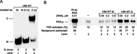
Dimers of the HDV ribozyme activate PKR. (A) Analytical native gel analysis of isolated and purified 1/99 WT monomer and dimer. Gel is 10% native PAGE run in 0.5× TBE. RNA monomers and dimers were first isolated as described in the Materials and Methods section “Purification of RNA monomers and dimers from native gels;” purification gel not shown. Then, certain monomer and dimer samples were added to an equal volume TEN200M20 (indicated with “s” for salt above the gel), followed by incubation at room temperature for 10 min. Prior to adding TEN200M20, certain monomer samples were heat renatured (indicated with “h” for heat above the gel) to remove possible dimer contaminants by incubating at 90°C for 1 min, followed by room temperature for 10 min, then 55°C for 10 min, followed by incubation at room temperature for 10 min. Both monomer-untreated (M) and monomer-treated with heat and salt (M (h+s)) are shown. Percent dimer is provided. This gel confirms that dimer identity has been largely retained and that, most importantly, monomer identity has been retained after native gel purification. (B) PKR activation by isolated and purified 1/99 WT HDV monomer and dimer. A no-RNA lane is provided, and phosphorylation activities are normalized to 0.01 μM 79-bp RNA. Both no-RNA and 79-bp RNA lanes contain 50 mM NaCl and 5 mM MgCl2. The no-RNA lane was subtracted from each lane in order to provide RNA-dependent activation values.
Absence of PKR activation by 1/99 WT monomer supports the inability of a globular RNA structure, confirmed in Figure 4, to activate PKR. In contrast, potent activation by the dimer RNA suggests that a helical structure can activate PKR, even at a concentration as low as 0.05 μM (Fig. 3B). Indeed, the optimal activation concentration of RNA has shifted at least 100-fold lower than in a heterogeneous mixture (cf. Figs. 2B and 3B). Even lower concentrations of HDV dimer were tested for PKR activation and resulted in less activation (Supplemental Fig. S1), once again revealing a bell-shaped dependence on RNA concentration and suggesting that 0.05 μM dimer is the optimal concentration for PKR activation. We previously noted that perfect 79-bp RNA optimally activates PKR between 0.01 and 0.1 μM (Zheng and Bevilacqua 2004). Thus, it appears that WT dimer interacts with PKR nearly as tightly as perfect 79-bp dsRNA. Overall, these findings support HDV ribozyme dimers, and not monomers, as being the activators of PKR. We also found that 1/99 Δ3 bp and 1/99 + 3 bp activated PKR as dimers but not monomers (Supplemental Fig. S2).
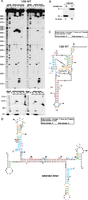
Enzymatic structure mapping of 1/99 WT HDV. (A) Structure mapping of 1/99 WT monomer and dimer. Monomeric and dimeric RNAs were prepared for structure mapping as described in the Materials and Methods section “Enzymatic structure mapping of RNA;” purification gel not shown. Sequencing lanes for G (T1) and all nucleotides (OH−) are provided in the left portion of each data set. The secondary structures were determined using RNases A (C, U-specific), T1 (G-specific), T2 (single-stranded-specific), and V1 (double-strand-specific). Residues 1–15 were resolved on a separate gel, shown at the bottom. (B) Analytical native gel analysis of RNAs used for structure mapping. Gel is 10% native PAGE run in 0.5× TBE. This gel confirms that monomer and dimer identity have been retained upon purification. (C) Monomer cleavage data superimposed on the monomer secondary structure are consistent with published HDV ribozyme structure, and include the noncanonical N1-N1, carbonyl-amino base pair between the terminal A and G of P4 (Ferre-D'Amare et al. 1998). (D) Dimer cleavage data superimposed on an extended dimer secondary structure model. In C and D, secondary structures are mapped with RNases A (orange), T1 (blue), T2 (green), and V1 (red). Closed and open triangles represent stronger and weaker cleavages, respectively. Most notably, L4 is largely protected in the dimer.
Secondary structure mapping of HDV monomers and dimers
The above experiments established a strong correlation between HDV ribozyme dimerization and PKR activation. To further our understanding of the molecular basis of PKR activation by HDV dimers, secondary structure mapping of monomer and dimer was performed (Fig. 4). Purified monomer and dimer RNA samples used for structure mapping retained their expected oligomeric identities on a native gel (Fig. 4B).
Enzymatic structure mapping of 1/99 WT monomer yielded a cleavage pattern in agreement with the published monomeric HDV ribozyme secondary structure (Been and Wickham 1997; Ferre-D'Amare et al. 1998), with L4 giving very intense single-stranded cleavages (Fig. 4A,C). The secondary structure data for 1/99 WT dimer, on the other hand, revealed the near absence of single-stranded cleavages in L4, consistent with the P4-kissing extended dimer depicted in Figure 4D. Residues A8 through C13 map differently for monomer and dimer, with more cleavage by the dsRNA-specific RNase V1 in monomer. This suggests that the dimer structure is not simply the native globular structure with a kissing P4 (Fig. 4D). The double-stranded cleavages observed in the monomer samples are reduced near G10 (RNase V1 cleavages migrate slower by ∼1 nt in this region due to leaving a 3′OH) (Brown and Bevilacqua 2005), consistent with J1/2 bend here. An absence of these RNase V1 cleavages in the dimer could result from A8 bulge and C13 mismatches depicted in the model in Figure 4D. This and other regions of cleavage superimposed on the secondary structure model in Figure 4D suggest that the PKR-activating HDV dimer is structurally distinct from the native globular HDV monomer, and is instead an extended dimer. It should also be noted that RNase V1 tends to cleave helical regions of at least 4–6 nt in length (Lowman and Draper 1986), which may account for the absence of V1 cleavage in the 44–47 bp of P4, which are also adjacent to tertiary structure, a bulged U, and have weak AU-rich pairing. Additionally, the more robust RNase T1—which is active in cleaving this RNA elsewhere—fails to cleave after any of the G's in the P4 region; thus, the P4 region in both the monomer and dimer is consistent with the helical structures shown (Fig. 4C,D). In addition, single-stranded cleavage of predicted double-stranded regions can occur at the last 2 bp of a helix, as observed in McGraw et al. (2009), which can account for patterns seen near G10, G25, and C26. Of the 24 strong hits observed, 19 are consistent with the proposed secondary structure, while the other five are in regions that may be due to breathing, allowing access to single-strand nucleases and stacking allowing access to RNase V1, which is known to cleave such regions (Lowman and Draper 1986). The potential for an ensemble of dimer species thus exists, but the data suggest that they would be closely related (see Conclusions).
Structure mapping of 1/99 Δ3 bp monomer and dimer was also performed, and monomer and dimer mapping was identical to 1/99 WT monomer and dimer, with extensive protection of L4 in the dimer, supporting generalization of the effects to this strong activator (Supplemental Fig. S3A–D). Lastly, we note that the extended dimer structures in WT RNA are 28 bp in length, which is below the minimal activating length of 33 bp (Manche et al. 1992; Zheng and Bevilacqua 2004), and which implicates helical contributions from the two adjacent 14-bp stem–loops. These three helical segments together would make a 56-bp dsRNA, which would be a strong PKR activator, as observed herein. The ability of multiple shorter helical fragments to promote activation has been noted previously in aptamers, pseudoknots, TAR, and the HCV IRES (Bevilacqua et al. 1998; Cohen-Chalamish et al. 2009; Shimoike et al. 2009; Heinicke 2010; Toroney et al. 2010). We note that similar activation of PKR by dimers of 1/99 Δ3 bp, WT, and +3 bp constructs at 0.1 μM RNA dimer (Supplemental Fig. S2B), supports the importance of regions outside of the 28-bp P4 dimerizing region, or else WT and especially +3 bp should activate more potently, given the above minimal activating length of 33 bp.
Lastly, we examined possible dimer folds using three secondary structure prediction programs: mfold v 3.5, RNAstructure, and pknotsRG (Zuker 2003; Reeder et al. 2007; Reuter and Mathews 2010). The bimolecular fold option was used for RNAstructure, while two copies of HDV with an A6 linker were used for mfold and pknotsRG. Without constraints, all three programs predict the extended kissing loop shown in Figure 4D as the lowest free energy structure, and are consistent with our dimer model. Our modeled stem–loop structures located on the 5′- and 3′-ends of this kissing region were present in most optimal and suboptimal structures.
CONCLUSIONS
In this study we examined the ability of the semiglobular HDV ribozyme to activate PKR. The ribozyme is mostly globular, but has a protruding 13-bp P4 pairing region. This region seemed the most likely one to promote activation of PKR, given that PKR is known to bind and be activated by long dsRNA. We thus examined PKR activation as a function of the length of P4 by preparing shortened and lengthened P4 variants. Surprisingly, 1/99 Δ3 bp was the most potent PKR activator, despite having fewer base pairs in the dimerized P4 region. It thus appears that the two adjacent 14-bp stem–loops contribute to activation, which was as high in magnitude as that of 79-bp dsRNA.
Correlation between extent of dimer formation, as determined by native PAGE, and activation of PKR was observed. Dimer formation was observed to be facilitated in the 1/99 Δ3-bp variant, which was likely due to destabilization of P4 upon removal of base pairs. To test whether dimer was the activating species, 1/99 WT and 1/99 Δ3 bp HDV monomer and dimer species were gently isolated from a native gel, confirmed to retain their original oligomeric states, and tested for activation of PKR. Monomers failed to activate PKR, while dimers were potent activators, activating PKR with affinity similar to 79-bp dsRNA. Structure mapping of 1/99 WT HDV monomer and dimers revealed that monomer folds as a native secondary structure of the ribozyme, but that the dimer species rearranges into an extended species consistent with the model in Figure 4D. Given the broad nature of the band on the native gels (e.g., Fig. 3A), it remains possible that there is an ensemble of related conformations in the dimer population.
The RNA dimer of the HDV ribozyme essentially doubles the number of contiguous base pairs, approaching the activating length, which is then likely exceeded by interaction with peripheral RNA regions. We observed a similar phenomenon in HIV TAR, where RNA dimerization drives PKR dimerization and activation (Heinicke et al. 2009). Indeed, most stem–loops dimerize at high concentration, owing to their self-complementary nature. Moreover, stem–loops can even polymerize, forming aggregates at high concentrations or with special sequences, which can activate PKR (Venkataraman et al. 2010). From the present study, it is clear that a semiglobular RNA can misfold into a dimer and activate the innate immune response protein, PKR. This contributes to the growing list of RNAs capable of activating PKR (Nallagatla et al. 2011) and provides a potential link between RNA misfolding and disease via innate immunity.
MATERIALS AND METHODS
Protein expression and purification
Full-length PKR containing an N-terminal (His)6 tag was cloned into pET-28a (Novagen, Inc.) and transformed into E. coli BL21(DE3) Rosetta cells (Novagen, Inc.) as previously described (Bevilacqua and Cech 1996; Zheng and Bevilacqua 2004; Nallagatla et al. 2007). Briefly, cells were sonicated and protein was purified by a Ni2+-agarose column (Qiagen, Inc.). A glutathione S-transferase (GST)–λ-Protein Phosphatase (PPase) fusion protein was cloned, expressed, and purified to treat partially phosphorylated PKR before PKR activation assays. λ-PPase was subcloned from pET21a containing λ-PPase and GST-PKR into pET-42a and transformed into E. coli BL21(DE3)pLysS cells. Protein was purified by sonication and filtration through a 0.45-micron filter, followed by GST column purification (Novagen). GST–λ-PPase was dialyzed into storage buffer: 50 mM HEPES (pH 7.5), 100 mM NaCl, 0.1 mM MnCl2, 0.1 mM EGTA, 2 mM DTT, 0.01% Brij 35, and 50% glycerol (New England Biolabs, NEB).
Dephosphorylated PKR was prepared by treatment with GST-λ-PPase using a standard NEB dephosphorylation protocol (Cohen and Cohen 1989). During dephosphorylation, GST-λ-PPase was present at ¼ the concentration of PKR. An additional glutathione column was used to separate PKR from GST–λ-PPase, and then PKR was dialyzed into a storage buffer of 10 mM Tris (pH 7.6), 50 mM KCl, 2 mM MgOAc, 10% glycerol, and 7 mM β-mercaptoethanol.
Plasmid and RNA preparation
HDV variants 1/99 Δ3 bp and 1/99 + 3 bp were prepared using QuikChange site-directed mutagenesis (Stratagene) to remove or introduce nucleotides in pUC19 plasmid containing −54/271 HDV sequence (accession number M28267, GenBank) (Makino et al. 1987; Diegelman-Parente and Bevilacqua 2002). Plasmids were linearized with BfaI for run-off T7 transcription (Ambion) (Chadalavada et al. 2000). Self-cleaved HDV RNAs were excised and soaked overnight at 4°C in TEN250 (10 mM Tris at pH 7.5, 1 mM EDTA, and 250 mM NaCl). Subsequently, RNAs were ethanol precipitated and resuspended in TE (10 mM Tris at pH 7.5 and 1 mM EDTA). Concentrations were determined spectrophotometrically. 5′-end-labeled RNAs were prepared by polynucleotide kinase treatment in the presence of [γ-32P]ATP.
Native gel analysis of HDV dimerization
Analytical native gel electrophoresis of RNA was performed for two reasons. The first was to analyze the monomeric and dimeric content of HDV ribozyme preparations (shown in Fig. 2A), and the other was to assess the retention of monomer and dimer formation after isolation of these species from preparative native gels (shown in Figs. 3A, 4B); see below for further details. Native gels contained 10% of 29:1 cross-linking polyacrylamide. The buffer both in the gel and for electrophoresis was 0.5× TBE (50 mM Tris base, 40 mM boric acid, and 0.5 mM EDTA) or THEM4 (34 mM Tris base, 66 mM HEPES, 0.1 mM EDTA, and 4 mM MgCl2). The buffer 0.5× TBE was used in most cases, as THEM4 and 0.5× TBE were found to have similar effects on the ratio of monomer to dimer. Native gels were run at 300 V and 16°C for 45 min prior to loading samples. In general, samples were fractionated for ∼2 h.
For the gel in which dimer and monomer content of non-native gel-purified RNAs were analyzed (Fig. 2A), trace amounts (∼1 nM) of 5′-end-labeled RNA was added to various concentrations of unlabeled RNA and renatured in TE by incubating at 90°C for 1 min, followed by incubating at 55°C for 10 min. In this experiment, samples in TE were then treated in one of three ways, by adding an equal volume to the following: (1) TE, (2) TEK400M20 (10 mM Tris at pH 7.5, 1 mM EDTA, 400 mM KCl, and 20 mM MgCl2), or (3) TEN400M20 (10 mM Tris at pH 7.5, 1 mM EDTA, 400 mM NaCl, and 20 mM MgCl2), followed by another incubation at room temperature for 10 min. Final concentrations in (2) and (3) were 200 mM KCl (or NaCl) and 10 mM MgCl2. In some cases, the final concentration of monovalent salt was decreased from 200 to 100 mM, as indicated in the main text. Prior to native gel analysis, 5% glycerol was added to the samples.
Confirmation of the upper and lower bands as dimer and monomer was achieved by comparison to 78-nt transcripts of tRNA dimer and monomers (Fig. 2A), prepared as described elsewhere (Wittenhagen and Kelley 2002; Roy et al. 2005). The 1/99 Δ3-bp WT, at 93 nt, is 15 nt or 19% longer than tRNA. We found that the monomer and dimer migrated ∼22% and 25% slower for HDV RNA than tRNA, which is in line with the 19% greater length of HDV RNA, with the P4 extension leading to less compaction and slightly slower mobility in HDV RNA than expected based on tRNA. Moreover, the dimer band only formed at high HDV RNA concentrations (Fig. 2A, cf. lanes 3–5 and lanes 6–8), confirming that it is a multimer. That the spacing between the monomer and dimer of tRNA (which is a 78-nt transcript) is nearly the same as the spacing between the monomer and dimer of 1/99 Δ3 bp (which is a 93-nt transcript) on the native gel (Fig. 2A) confirms that the upper band of HDV RNA is a dimer. A kissing loop complex with protection of L4 further confirms this assignment.
Native gels were also run to assess retention of the monomeric and dimeric states. Analytical gels are shown in Figures 3A and 4B and generally3 omitted the high-temperature renaturation in an effort to preserve any dimer, and were exposed to a storage screen for 16 h and analyzed on a PhosphorImager (Molecular Dynamics).
Purification of RNA monomers and dimers from native gels
Native gels were used to prepare monomer and dimer RNA. In most cases, 20 or 50 μM RNA in TE was renatured in the presence of radiolabeled RNA by incubating at 90°C for 1 min, followed by incubation at 55°C for 10 min. In some cases, 1/99 WT and 1/99 + 3-bp RNA dimers were prepared by incubating in TEN100 at 95°C for 4 min, followed by slow cooling to 40°C over 1.5 h (Supplemental Fig. S2). Next, 5% glycerol was added to the RNA, and it was fractionated on a 0.5× TBE native gel for 2 h. Monomer and dimer RNA bands were visualized by UV shadowing and excised. RNAs were crushed and soaked overnight in TEN250 at 4°C, ethanol precipitated the next day, resuspended in TE, and stored at −20°C. All radiolabeled samples were refractionated on an analytical native gel, as described above, to assess retention of monomer and dimer state.
PKR activation assays
RNAs were tested for their ability to activate PKR, as shown in Figures 2B and 3B. GST-λ-PPase-treated PKR (0.6 μM) was incubated with various concentrations of RNA, in a final concentration of 20 mM HEPES (pH 7.5), 5 mM MgCl2, 50 or 100 mM NaCl, 1.5 mM DTT, 100 μM ATP (Ambion), and 15 μCi [γ-32P]ATP. Heterogeneous RNA species, which are a mixture of monomer and dimer species, were prepared by incubating 4x concentration RNA at 90°C for 1 min, followed by 55°C for 10 min (e.g., Fig. 2B). RNAs that were isolated from a native gel as monomer or dimer (as described in the previous subsection) were subjected to this 90°C step (e.g., Figs. 3A, 4B) only for monomer preparations. For certain RNAs, an equal volume of TEN400M20 or TEN200M20 was added, followed by incubation at room temperature for 10 min to give 2× stocks that were then added to an appropriate PKR reaction mixture.
Prior to activation assays, RNA isolated as monomers was renatured briefly at 90°C to help remove any dimer and retain pure monomeric identity as previously described (Heinicke et al. 2009), and the final concentrations of RNA did not exceed 0.6 μM to avoid the increased the likelihood of introducing small amounts of dimer into renatured monomer samples; RNAs isolated at dimers were not heated at this step. PKR activation reaction mixtures were incubated at 30°C for 10 min, quenched with SDS loading buffer, and loaded on 10% SDS-PAGE (Pierce). Gels were exposed to a storage PhosphorImager screen, and intensities of the PKR bands were quantified using a PhosphorImager (Molecular Dynamics). In activation assay gels, a no-RNA lane is provided, and phosphorylation activities are normalized to 0.01 μM 79-bp RNA. Fold-effects are reported for relative activation after the no-RNA background was subtracted.
Enzymatic structure mapping of RNA
RNA monomers were renatured at 3 μM in water by incubating at 90°C for 3 min to remove any dimer, while dimer samples were not renatured (Heinicke et al. 2009). Final concentrations of monomer and dimer RNAs for both native and denaturing structure mapping reactions were 1 and 0.5 μM, respectively, to give the same total concentration of nucleotides, which allowed the same amount of ribonuclease to be used for mapping monomeric and dimeric RNAs. Ribonuclease concentrations were 4 ng/mL RNase A, 0.001 U/μL RNase T1, 5 × 10⁻⁴ U/μL RNase T2, and 0.002 U/μL RNase V1. Higher concentrations of ribonucleases were tested, but resulted in nonspecific cleavage. Ribonucleases were diluted into 10 mM Tris (pH 8.0), 10% glycerol, and 1 mM DTT as needed. Native RNA cleavage reactions were performed in 20 mM MES (pH 6.0), 50 mM NaCl, and 10 mM MgCl2 at 37°C for 15–60 min. Denaturing RNA cleavage reactions, used as sequencing lanes, were 0.1 U/μL T1, 4.7 M urea, 14 mM sodium citrate (pH 3.5), and 0.7 mM EDTA at 50°C for 15 min, and hydrolysis reactions were 50 mM Na2CO3/NaHCO3 (pH 9.0) and 1 mM EDTA at 90°C for 4 min. Fractionated samples were loaded onto 12% PAGE/TBE/8.3 M urea denaturing gel.
SUPPLEMENTAL MATERIAL
Supplemental material is available for this article.
ACKNOWLEDGMENTS
Special thanks to Durga Chadalavada for assistance with HDV preparation. We also thank Durga Chadalavada and Chelsea Hull for helpful comments on the manuscript. This work was supported by National Institutes of Health grant GM-58709 (P.C.B.).
Footnotes
-
↵2 Corresponding author
E-mail pcb5{at}psu.edu
-
↵3 The only exception was certain lanes desired to be monomer, such as Fig. 3A, lane 2, in which a renaturation was conducted.
-
Article published online ahead of print. Article and publication date are at http://www.rnajournal.org/cgi/doi/10.1261/rna.034744.112.
- Received June 5, 2012.
- Accepted September 20, 2012.
- Copyright © 2012 RNA Society




