


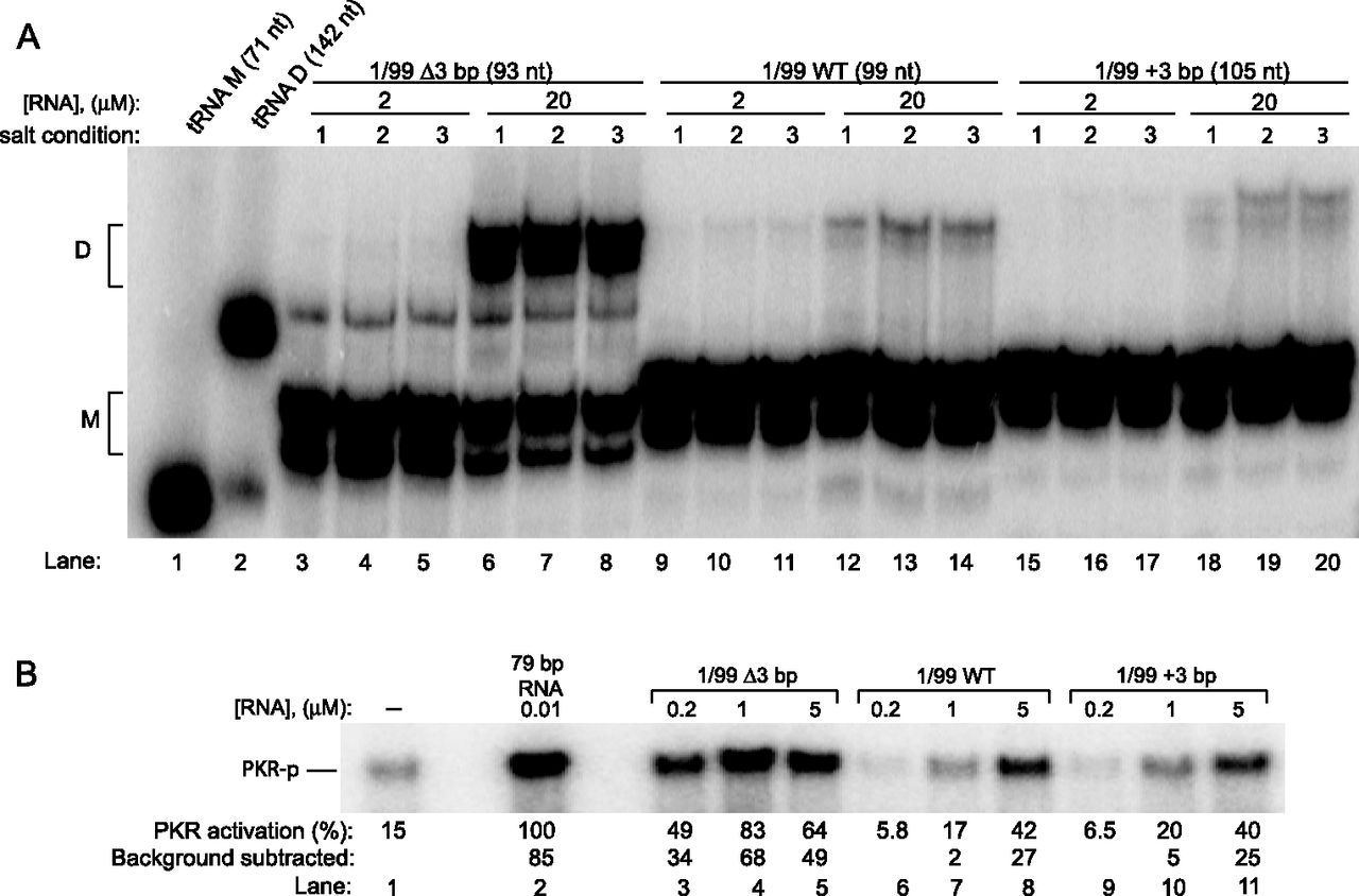
Native gel analysis of HDV dimerization and its association with PKR activation. (A) Native gel analysis of RNA. Gel is 10% native PAGE run in THEM4. 1/99 Δ3 bp, WT, and +3 bp were renatured at 2 or 20 μM concentrations with trace 5′-end labeled RNA in TE, by incubating at 90°C for 1 min, followed by room temperature for 10 min, then 55°C for 10 min, followed by incubation at room temperature for 10 min. The sample was then mixed with an equal volume of one of the following salt conditions (labeled in gel): (1) TE, (2) TEK400M20, or (3) TEN400M20; final ionic concentrations in 2 and 3 were 200 mM KCl (or NaCl) and 10 mM MgCl2. Number of nucleotides in cleaved monomer HDV is provided above the gel. The faster migrating bands are assigned as monomer (M) and the slower migrating bands as dimer (D) as described in the Materials and Methods; assignment of these bands is facilitated by structure mapping (see below) and by comparison to lanes 1 and 2, which are tRNA monomer (M) and dimer (D), with the dimer prepared as per Wittenhagen and Kelley (2002) and Roy et al. (2005). (The minor, faster-mobility band associated with M and D was not always present in native gels [e.g., absent in Figs. 3A, 4B] and may represent alternative RNA structures induced by the presence of divalent salt in THEM4 buffer, which is absent in Figs. 3A, 4B.) (B) PKR activation by HDV variants containing a mixture of the M and D species. Renaturation and ionic conditions are provided in the Materials and Methods. A no-RNA lane is provided, and phosphorylation activities are normalized to 0.01 μM 79 bp RNA. Both no-RNA and 79-bp RNA lanes contain a final concentration of 100 mM NaCl and 5 mM MgCl2. The no-RNA lane was subtracted from each lane in order to provide RNA-dependent activation values.




