


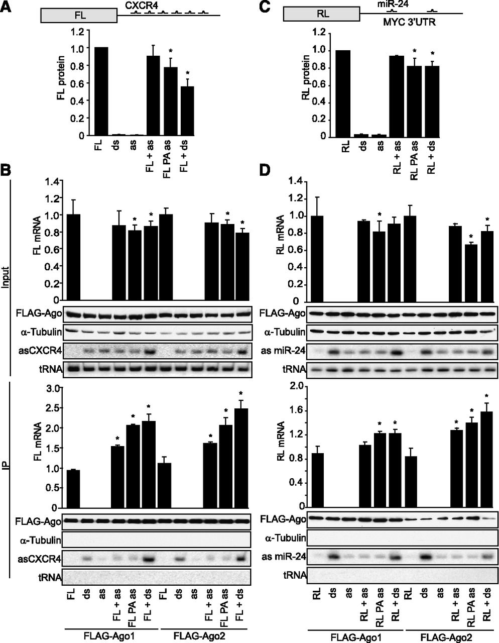
Ago1 and Ago2 bind and repress preannealed miRNA–mRNA duplexes in cells. (A) Preannealed artificial mRNA–miRNA duplexes are repressed in cells. HeLa cells were transfected with in vitro transcribed, capped, and polyadenylated FL mRNA containing six imperfect CXCR4 binding sites in its 3′ UTR (FL6X; top) and with antisense (as), preannealed (PA), or double-stranded (ds) CXCR4. After 16 h, luciferase assays were performed and normalized to untargeted Renilla luciferase control. (B) Ago1 and Ago2 are recruited to FL6X preannealed to CXCR4. HeLa cells were transfected with constructs expressing Flag-Ago1 or Flag-Ago2, and 24 h later, FL6X was transfected along with indicated forms of the CXCR4 miRNA. After 16 h, anti-Flag immunoprecipitation (IP) was performed and verified by Western blotting for Flag and α-tubulin. CXCR4 in the input and IP fractions was detected by Northern blotting and normalized to tRNA, and FL mRNA was detected by RT-qPCR and normalized to GAPDH. (C) Preannealed endogenous target mRNA–miRNA duplexes are repressed in cells. A549 cells were transfected with in vitro transcribed, capped, and polyadenylated Renilla (RL) mRNA containing MYC 3′ UTR with two imperfect miR-24 binding sites in its 3′ UTR (RL-MYC 3′ UTR; top) and with antisense (as), preannealed (PA), or double-stranded (ds) miR-24. After 16 h, luciferase assays were performed and normalized to untargeted Renilla luciferase control. (D) Ago1 and Ago2 are recruited to RL-MYC 3′ UTR preannealed to miR-24. A549 cells were transfected with constructs expressing Flag-Ago1 or Flag-Ago2, and 24 h later, RL-MYC 3′ UTR mRNA was transfected along with indicated forms of miR-24. After 16 h, anti-Flag G IP was performed and verified by Western blotting for Flag and α-tubulin. MiR-24 in the input and IP fractions was detected by Northern blotting and normalized to tRNA, and RL mRNA was detected by RT-qPCR and normalized to GAPDH. Bar graphs, mean ± SD from three independent experiments. (*) P < 0.05; Student's t-test.




