


Biogenesis of telomerase ribonucleoproteins
- Department of Molecular and Cell Biology, University of California at Berkeley, Berkeley, California 94720-3200, USA
Abstract
Telomerase adds simple-sequence repeats to the ends of linear chromosomes to counteract the loss of end sequence inherent in conventional DNA replication. Catalytic activity for repeat synthesis results from the cooperation of the telomerase reverse transcriptase protein (TERT) and the template-containing telomerase RNA (TER). TERs vary widely in sequence and structure but share a set of motifs required for TERT binding and catalytic activity. Species-specific TER motifs play essential roles in RNP biogenesis, stability, trafficking, and regulation. Remarkably, the biogenesis pathways that generate mature TER differ across eukaryotes. Furthermore, the cellular processes that direct the assembly of a biologically functional telomerase holoenzyme and its engagement with telomeres are evolutionarily varied and regulated. This review highlights the diversity of strategies for telomerase RNP biogenesis, RNP assembly, and telomere recruitment among ciliates, yeasts, and vertebrates and suggests common themes in these pathways and their regulation.
Keywords
- ribonucleoprotein biogenesis
- telomerase RNA
- telomerase reverse transcriptase
- telomerase holoenzyme
- telomere maintenance
INTRODUCTION
The evolution of linear chromosomes in the common ancestor of eukaryotes occurred despite several challenges posed by this mode of genome organization. First, conventional primer-requiring DNA polymerases cannot copy the 3′ end of a DNA template, leading to the progressive loss of end sequence with every round of DNA replication. Second, chromosome ends resemble double-stranded DNA breaks whose recognition and repair results in chromosome fusions. Third, DNA ends are vulnerable to the destructive action of exonucleases. A solution to these challenges was to form a protective cap structure from a tandem array of telomeric repeats with a net length of tens to thousands of base pairs (Gomes et al. 2010; Jain and Cooper 2010 O'Sullivan and Karlseder 2010). Telomeric DNA termini typically have a 3′ overhang, which can invade the duplex to form a loop that sequesters the single-stranded end. Single-stranded and double-stranded DNA-binding proteins coat the telomeric repeat DNA to assemble an end-capping structure (de Lange 2010; Stewart et al. 2012).
Telomere maintenance in most eukaryotes depends on new repeat synthesis by the specialized reverse transcriptase (RT) telomerase (Blackburn et al. 2006). Telomerase is a ribonucleoprotein (RNP) enzyme with two catalytically essential subunits: the telomerase reverse transcriptase protein (TERT) and the telomerase RNA (TER). TERT copies a template within TER to determine the repeat sequence, and outside of the active site additional domains of both subunits contribute to synthesis (Blackburn and Collins 2011). Many telomerase enzymes maintain productive binding to the substrate DNA during synthesis across the entire template (nucleotide addition processivity) and also during multiple rounds of template copying (repeat addition processivity, RAP). The processivity and fidelity of repeat synthesis vary across telomerases from different organisms and even telomerase from the same organism under different growth conditions (Chang et al. 2007; Collins 2009; Zhao et al. 2011). It is possible that some TERT proteins do not copy an internal template, perhaps reflected in the evolutionary loss of entire TERT domains (Blackburn and Collins 2011). Other organisms including Drosophila have lost TERT and TER entirely and instead use recombination- or transposition-based mechanisms for telomere maintenance (Pardue and DeBaryshe 2011).
In general, TERTs contain four domains: the telomerase “essential” N-terminal (TEN) domain, the telomerase RNA-binding domain (TRBD), the RT domain, and the C-terminal extension (CTE). The TEN domain traps single-stranded telomeric DNA and also interacts with TER (O'Connor et al. 2005; Jacobs et al. 2006; Robart and Collins 2011). The single-stranded DNA-handling function of the TEN domain likely promotes processive repeat synthesis by capturing the substrate and maintaining association with the single-stranded product (Collins 2011). The TRBD confers the specificity of interaction between TERT and TER (Lai et al. 2001; O'Connor et al. 2005). In the RT domain, motifs conserved in the evolutionarily related retrotransposon RTs form the active site, where aspartic acid residues coordinate the magnesium ions necessary for catalysis of dNTP addition (Lingner et al. 1997b; Belfort et al. 2011). In TERTs, this domain also positions the template and aligns the substrate 3′ end (Xie et al. 2010; Qi et al. 2012). The CTE may enhance nucleic acid association and/or otherwise contribute to RT domain function (Autexier and Lue 2006; Wyatt et al. 2010). A crystal structure of TERT from the flour beetle Tribolium castaneum, which lacks a TEN domain, revealed CTE contacts with the TRBD that create a protein ring around a model RNA–DNA hybrid (Gillis et al. 2008; Mitchell et al. 2010). It is possible that the CTE influences the telomerase catalytic cycle by affecting the conformation of this ring. Further insights will be greatly aided by high-resolution structure of TERT domain complexes with an endogenous TER.
TERs vary more than TERTs in their folded domain organization, with disparate sequences and sizes ranging from about 150 nucleotides (nt) in ciliates to more than 10 times that length in some yeasts. However, functional assays suggest that several TER elements may be considered shared between ciliates, yeasts, and vertebrates, as illustrated in Figure 1 and reviewed extensively elsewhere, and therefore not individually referenced below (Theimer and Feigon 2006; Collins 2009; Blackburn and Collins 2011; Zhang et al. 2011; Podlevsky and Chen 2012). First is the single-stranded template, whose complement typically corresponds to about 1.5 repeats of the telomeric DNA sequence. Notable exceptions to this template length occur in yeasts with long and/or degenerate telomeric repeat sequences (Lue 2010). The 3′ region of the template aligns the DNA substrate, while the 5′ portion is copied. Adjacent to the template is a unique pseudoknot fold stabilized by triple-helix formation. Despite its structural conservation and importance for TER folding overall, the specific function of the TER pseudoknot remains an outstanding question. Based on studies to date, we suggest that a unifying model for pseudoknot function would be in positioning the template relative to the active site and other TER and TERT domains.
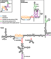
Diagram of TER secondary structures highlighting functional motifs. The template, pseudoknot, TBE, and STE are common to ciliate, yeast, and vertebrate TERs. The STE is distal stem–loop IV in T. thermophila, conserved region 4/5 (CR4/5) in human, and a three-way helix junction in yeasts. Species-specific RNP stability elements recruit p65 in T. thermophila, H/ACA proteins in human, and Sm proteins in yeasts (also Ku in Saccharomyces). The binding sites for holoenzyme proteins that do not affect RNA stability, namely Est1 in yeasts and the CAB box-binding protein WDR79/TCAB1 in humans, are indicated. T. thermophila TER also contains a template recognition element (TRE) that contributes to template utilization.
The template-pseudoknot domain is typically closed by long-range base-pairing of sequence at or near the TER 5′ end (Fig. 1). This stem or a separate hairpin immediately 5′ of the template can provide a 5′ template boundary element (TBE) to prevent copying of nontemplate TER sequence. Separate from all of the other conserved TER elements described above is a “stem terminus” element (STE), comprised of either a terminal hairpin, a hairpin from a three-way junction, or a three-way junction alone (Fig. 1). The STE stimulates telomerase activity, at least in part, through direct interaction with the TERT TRBD. Because some STE sequence substitutions reduce activity without an evident change in TERT binding, the STE motif is likely to have an additional role in RNP folding and/or catalytic activation. One hypothesis for this additional role is an allosteric influence on TERT and/or TER domain conformation.
Cellular telomerase holoenzymes are multisubunit complexes that fractionate by gel filtration with an apparent mass of 500 kDa or more (Collins 2009). However, only TERT and TER are required to reconstitute telomerase catalytic activity on oligodeoxynucleotide substrates in vitro (Weinrich et al. 1997). Beyond the TER motifs that bind TERT and orchestrate the telomerase catalytic cycle, TERs possess additional motifs that recruit proteins involved in TER processing and RNP stability or localization in vivo. In addition, other telomerase holoenzyme proteins direct and regulate physical interactions with telomeric chromatin and functional engagement with the chromosome 3′ end. The identities of telomerase holoenzyme proteins and their exact mechanisms of function differ between organisms, but some of their biochemical roles bear striking similarities. This review aims to describe the unique and shared features of telomerases from ciliates, yeasts, and vertebrates with an emphasis on pathways of telomerase RNA biogenesis and RNP assembly. Telomerase physical recruitment to, elongation of, and regulation at telomeres are modulated by dynamic telomeric chromatin reviewed extensively elsewhere (Stern and Bryan 2008; Moser and Nakamura 2009; Schoeftner and Blasco 2009; de Lange 2010; Stewart et al. 2012). Most telomerase studies have involved Tetrahymena thermophila, Saccharomyces cerevisiae, Schizosaccharomyces pombe, mice, or cultured human cells. Less-intensively studied but important additional model systems include other ciliates, yeasts, vertebrates and also plants (Collins 1999; Teixeira and Gilson 2005; Watson and Riha 2010; Cifuentes-Rojas et al. 2011; Yu 2012).
CILIATE TELOMERASE
Ciliate telomerase RNA structure
Ciliate TERs are the smallest to be characterized to date. The model laboratory ciliate, T. thermophila, possesses a 159-nt TER (Greider and Blackburn 1989) that includes all of the conserved motifs summarized above (Fig. 1). The 9-nt template is flanked at its 5′ end by a TBE and at its 3′ end by a pseudoknot. These three motifs are enclosed by long-range base-pairing of stem I (Fig. 1). In addition to defining the template boundary, the TBE and its adjacent single-stranded regions also provide the high-affinity TERT-binding site (Autexier and Greider 1995; Lai et al. 2002). A recent single-molecule fluorescence resonance energy transfer study found that the T. thermophila TER pseudoknot remains stably folded throughout the catalytic cycle (Mihalusova et al. 2011). Remarkably, the pseudoknot can form even when some of the stem pairing is disrupted (Mihalusova et al. 2011), likely accounting for the modest impact of some, but not other, pseudoknot-destabilizing mutations on telomerase holoenzyme assembly and activity (Gilley and Blackburn 1999; Cunningham and Collins 2005).
In ciliate TERs, terminal stem IV and its loop comprise the STE (Fig. 1). Nucleotides within loop IV contribute to TERT binding in a manner important for RNP conformational stability in vitro and biological stability in vivo (Stone et al. 2007; Robart et al. 2010). T. thermophila TER also has another motif immediately 3′ of the template termed the template recognition element (TRE) (Fig. 1), which contributes to efficient copying through mid-template positions and improves the in vitro use of oligonucleotide templates physically separate from the rest of TER (Miller and Collins 2002; Cunningham and Collins 2005). Curiously, cells expressing a TER with TRE sequence substitutions have longer than normal telomeres, perhaps linked to altered enzyme pausing during repeat synthesis (Cunningham and Collins 2005).
Ciliate telomerase RNP maturation and TERT–TER assembly
Ciliate telomerase RNP assembly begins with TER transcription by RNA polymerase III (Greider and Blackburn 1989). Mature TER retains the 3′ uridine-rich termination sequence of the primary transcript (Fig. 2). In T. thermophila the TER 3′ polyuridine tail along with stems I and IV provides a binding site for the telomerase-specific p65 protein (Fig. 2). This La-family telomerase holoenzyme protein is required for TERT and TER accumulation in vivo (Witkin and Collins 2004; O'Connor and Collins 2006). Here, we will use the designation “RNP catalytic core” to describe the physiologically assembled minimal catalytically active RNP (for example, the p65–TER–TERT ternary complex), which includes subunits essential for TER folding and stability in vivo but not for optimized TERT + TER minimal RNP reconstitution in vitro. The interaction of p65 with TER stabilizes a kink in stem IV necessary for tight TERT binding (Stone et al. 2007; Singh et al. 2012). Binding of p65 also induces additional conformational changes in stem–loop IV and beyond that promote TERT assembly and catalytic activity (Prathapam et al. 2005; Richards et al. 2006; Berman et al. 2010; Akiyama et al. 2012; Singh et al. 2012).
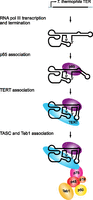
T. thermophila telomerase RNP biogenesis. T. thermophila TER is transcribed by RNA polymerase III. The binding of p65 stabilizes a kink in stem IV that optimally positions loop IV for TERT binding. The p65-TER–TERT ternary complex then interacts with a complex of p75, p50, p45, and p19 that recruits the single-stranded telomeric DNA-binding protein Teb1.
Bridging the ciliate RNP catalytic core to telomere substrates
The T. thermophila telomerase holoenzyme has been purified to homogeneity, enabling extensive subunit characterization. Beyond the RNP catalytic core, five associated proteins designated p19, p45, p50, p75, and Teb1 (Fig. 2) are required for telomere maintenance in vivo (Witkin and Collins 2004; Witkin et al. 2007; Min and Collins 2009). Four of these five proteins (p19, p45, p50, and p75) contribute to the telomere adaptor subcomplex (TASC) that bridges the RNP catalytic core to Teb1 (Min and Collins 2009; Eckert and Collins 2012). Teb1 joining generates the complete telomerase holoenzyme, which in T. thermophila retains a remarkably stable grip on product DNA and thus supports high RAP in vitro (Greider 1991).
Teb1 binds directly and with high affinity to single-stranded telomeric repeat DNA (Min and Collins 2009, 2010; Zeng et al. 2011). Although Teb1 has an oligonucleotide/oligosaccharide (OB) fold architecture homologous to the large subunit of Replication Protein A (RPA), it is telomerase-specific in its physical interactions and sequence-specific in its recognition of tandem T2G4 repeats. Teb1 stimulation of RAP in vitro results in part from activating protein–protein interaction(s) and in part from its binding to nascent product DNA (Min and Collins 2010), which could be important in vivo independent of elongation RAP per se. Because the Teb1 and p50 subunits are extensively proteolyzed and/or substoichiometric in the pool of holoenzyme purified from cell extract (Min and Collins 2009), a heterologous system for reconstitution of recombinant holoenzyme will be required to fully understand the roles of TASC and Teb1 in telomere elongation. T. thermophila telomerase physical recruitment to and/or functional activation at telomeres may also involve the telomere-associated Pat1 protein, whose depletion causes gradual telomere shortening without compromised end-protection (Linger et al. 2011).
YEAST TELOMERASE
Yeast telomerase RNA structure
Yeast telomerase RNAs are relatively large (1000 nt or more) with a template, TBE, and pseudoknot brought together by long-range base-pairing of stem I (Fig. 1). In the S. cerevisiae TER, TLC1, a high-affinity binding site for the TERT protein Est2 is within this conserved region (Livengood et al. 2002; Zappulla et al. 2005). Most of yeast TER length derives from three long stems, or “arms,” extending from the conserved central region (Fig. 1). One of these arms is positioned immediately 5′ of the template and provides a TBE (Tzfati et al. 2000; Seto et al. 2003; Box et al. 2008b). In addition, in Saccharomyces but not other yeast species, the terminal stem–loop of this arm forms a binding site for the Ku heterodimer (Fig. 1), which enhances in vivo RNP accumulation, nuclear localization, and telomere recruitment (Peterson et al. 2001; Stellwagen et al. 2003; Fisher et al. 2004; Gallardo et al. 2008; Mozdy et al. 2008). The second RNA arm occurs between the template and pseudoknot and contains a binding site for the regulatory protein Est1 (Fig. 1), which together with another holoenzyme subunit, Est3, allows telomere elongation in vivo (see below and for review Osterhage and Friedman 2009; DeZwaan and Freeman 2010). A similar motif in the S. pombe TER, TER1, interacts with an Est1 ortholog (Webb and Zakian 2012), but no Est3 ortholog is known to exist in this species.
The third RNA arm is the extremely long stem I, extended by a hairpin formed by fold-back of the 3′ end (Fig. 1). This 3′ terminal hairpin contains the STE as a three-way junction motif that stimulates catalytic activity and may contact TERT in Kluyveromyces lactis and likely other yeasts (Brown et al. 2007). A single-stranded uridine-rich motif near the 3′ end of yeast TERs binds to Sm proteins (Fig. 1), which contribute to the maturation and stability of the RNA (Seto et al. 1999; Tang et al. 2012). Studies of TLC1 have demonstrated that the RNA arms can be truncated substantially and still support activity in vitro or prevent senescence in vivo (Zappulla et al. 2005), giving rise to the impression of TER as a conformationally flexible scaffold for protein assembly (Zappulla and Cech 2006). However, minimized TLC1 accumulates to levels much lower than wild type, maintains short telomeres, and reduces fitness (Zappulla et al. 2005).
Studies of yeast TERs have provided insights into the function of the pseudoknot. In K. lactis TER, some pseudoknot sequence substitutions prevent copying of the full template (Tzfati et al. 2003). Another study used fragments of TLC1 assembled with Est2 in rabbit reticulocyte lysate to determine that disruption of the pseudoknot triple helix reduced telomerase activity but not Est2 binding (Qiao and Cech 2008). A role for the pseudoknot in substrate DNA positioning was proposed based on the crosslinking of pseudoknot nucleotides within a minimal recombinant RNA alone to the 3′ end of a telomeric DNA oligonucleotide (Qiao and Cech 2008).
Yeast telomerase RNP maturation and TERT–TER assembly
Studies of yeast telomerase RNA biogenesis have focused on S. cerevisiae TLC1 and more recently also on S. pombe TER1. Both are independently transcribed by RNA polymerase II and accumulate primarily as unpolyadenylated species (Figs. 3, 4) with a minor fraction (5%–10%) occurring in a polyadenylated form (Chapon et al. 1997; Leonardi et al. 2008). Telomerase holoenzyme contains the unpolyadenylated form (Bosoy et al. 2003; Leonardi et al. 2008).
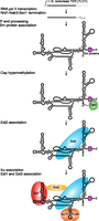
S. cerevisiae telomerase RNP biogenesis. S. cerevisiae TLC1 is transcribed by RNA polymerase II, and transcription is terminated by the Nrd1–Nab3–Sen1 pathway. The 3′ end is processed by the nuclear exosome and TRAMP complex, leaving a short adenosine-rich tail that is ultimately removed. Sm protein binding near the 3′ end stabilizes the RNA and recruits the cap hypermethylase Tgs1, which modifies the 5′ cap to TMG, indicated by a diamond. The TERT subunit Est2 binds directly to TLC1. The Ku heterodimer recognizes a distinct binding site on TLC1 to promote RNP accumulation and nuclear import. The regulatory subunit Est1 can bind directly to TLC1 and with Est3 stimulates telomerase function at telomeres.
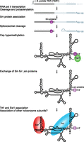
S. pombe telomerase RNP biogenesis. S. pombe TER1 is transcribed by RNA polymerase II as a precursor that includes a downstream intron and exon. TER1 secondary structure has yet to be experimentally validated; it is illustrated here as similar to other yeast TERs. Sm proteins assemble on the precursor near the mature TER1 3′ end. Spliceosomal cleavage at the 5′ splice site releases mature TER1, which escapes ligation to the downstream exon. Sm proteins then recruit the cap hypermethylase Tgs1, which modifies the 5′ cap to TMG, indicated by a diamond. Sm proteins are then replaced by Lsm proteins that protect the 3′ end from degradation, followed by assembly of Est1 and the TERT subunit Trt1.
Although previously suggested to be a precursor, recent results suggest that the polyadenylated form of TLC1 is not an intermediate in the accumulation of the unpolyadenylated mature form (Noël et al. 2012). The 3′ end of functional TLC1 is produced by the Nrd1–Nab3–Sen1 pathway (Fig. 3), which also matures the transcripts of small nuclear (sn) and small nucleolar (sno) RNAs in yeast (Kuehner et al. 2011; Noël et al. 2012). In this pathway, specific patterns of RNA polymerase II C-terminal domain phosphorylation recruit the RNA-binding proteins Nrd1 and Nab3, which recognize sequences in the precursor transcript downstream from the mature RNA 3′ end. They interact with the Sen1 helicase, which is thought to terminate transcription by unwinding the RNA–DNA hybrid in the RNA polymerase active site. The TRAMP (Trf4, Air2, Mtr4) complex then adds a short polyadenosine tail to the 3′ end (Fig. 3), which is ultimately removed by the nuclear exosome (Jamonnak et al. 2011; Kuehner et al. 2011).
The processing pathway that generates mature S. pombe TER1 is surprisingly different from that used by S. cerevisiae TLC1 (Fig. 4). The 3′ extended, polyadenylated form of TER1 does appear to serve as the precursor for mature TER1 3′ end formation (Box et al. 2008a). An intron following the exon encoding mature TER1 is recognized by the spliceosome, and cleavage occurs at the 5′ splice site without ligation (Fig. 4), an outcome potentially favored by a weak 3′ splice-site sequence and the atypical length of sequence between this site and the branch point (Box et al. 2008a). The same mechanism may generate the 3′ end of Candida TERs, which exhibit a similar conservation of 5′ splice site and branch point sequences (Gunisova et al. 2009).
TLC1 and TER1, like snRNAs, assemble with Sm proteins and acquire a 2,2,7-trimethylguanosine (TMG) cap (Figs. 3, 4). The Sm proteins SmB, SmD1, SmD2, SmD3, SmE, SmF, and SmG form a heteroheptameric ring that binds a single-stranded uridine-rich region at the 3′ ends of TLC1 and TER1, promoting RNP accumulation and also TER cap hypermethylation via a protein–protein interaction with the TMG synthase Tgs1 (Seto et al. 1999; Leonardi et al. 2008; Tang et al. 2012). Kluyveromyces and Candida TERs conserve this sequence element and thus likely also Sm protein interactions (Gunisova et al. 2009). In S. pombe but not S. cerevisiae, the Sm ring on TER is replaced by a related Lsm2–8 complex that promotes the association of TER1 with the S. pombe TERT protein, Trt1 (Fig. 4), and protects the mature TER1 3′ end from exonuclease activity (Tang et al. 2012).
Studies of yeast TER subcellular trafficking have primarily investigated S. cerevisiae TLC1. TLC1 cap hypermethylation occurs in the nucleolus (Mouaikel et al. 2002). TMG-capped TLC1 is then exported to the cytoplasm via the Crm1 export complex and is later reimported via Mtr10 and/or Kap122 importins (Ferrezuelo et al. 2002; Gallardo et al. 2008). In S phase, TLC1 can be detected at a few clustered telomeres (Gallardo et al. 2011). The findings that all three telomerase holoenzyme Est proteins are required for endogenous TLC1 nuclear localization, and that overexpressed Est1 or Est2 localizes to the nucleus, suggest the possibility that these proteins assemble on TLC1 in the cytoplasm and facilitate its nuclear reentry (Teixeira et al. 2002; Gallardo et al. 2008). However, unlike the case for T. thermophila TER, the biological accumulation of yeast TERs does not require TER assembly with TERT (Lingner et al. 1997a).
Bridging the yeast RNP catalytic core to telomere substrates
S. cerevisiae Est1 and Est3 are required for telomere elongation in vivo but not for RNP catalytic core activity in vitro (Lingner et al. 1997a). It was thus proposed that these proteins physically recruit the RNP to telomeres or subsequently activate it for telomere elongation. In addition to binding TLC1 (Fig. 3), S. cerevisiae Est1 interacts with the single-stranded telomeric DNA-binding protein Cdc13, which alternatively can form a telomere-associated RPA-like complex with Stn1 and Ten1 designated CST (Qi and Zakian 2000; Grandin et al. 2001; Pennock et al. 2001; Gao et al. 2007; Wu and Zakian 2011). Many studies have characterized the interaction of Est1 and Cdc13 as a physical bridge of the telomerase RNP catalytic core to a telomere substrate, as thoroughly reviewed elsewhere (Osterhage and Friedman 2009; DeZwaan and Freeman 2010). Although Est2 is detected at telomeres by chromatin immunoprecipitation (ChIP) throughout the cell cycle, Est1 is not telomere-associated during G1 when it is degraded by the proteasome (Taggart et al. 2002; Osterhage et al. 2006). These and other findings have led to the suggestion that in addition to physically bridging a telomerase RNP to a telomere, Est1 also stimulates S. cerevisiae telomerase catalytic activity as part of its biological function (DeZwaan and Freeman 2010).
Recent studies have begun to define the biochemical role(s) of Est3 in yeast telomerase holoenzymes. Candida albicans Est3 stimulates in vitro elongation of some oligonucleotide primers, and Saccharomyces castelli or S. cerevisiae Est3 stimulates catalytic activity overall (Hsu et al. 2007; Lee et al. 2010; Talley et al. 2011). Est3 interacts directly with the Est2 TEN domain in S. cerevisiae (Fig. 3) and S. castelli (Lee et al. 2010; Talley et al. 2011). A study of recombinant Est3 from yeast lacking a known Est1 demonstrated crosslinking to single-stranded telomeric DNA dependent on Est3 interaction with the Est2 TEN domain (Yen et al. 2011). Incorporation of Est3 into the S. cerevisiae or C. albicans telomerase holoenzyme has a variably reported dependence on Est1 (Osterhage et al. 2006; Hsu et al. 2007; Lee et al. 2010; Tuzon et al. 2011).
S. pombe telomerase holoenzyme assembly differs from that of S. cerevisiae in Est protein association (Fig. 4). Reciprocally with some other characterized yeasts, S. pombe has Est1, but not an identified Est3 (Beernink et al. 2003). S. pombe Est1 has been proposed to recruit telomerase to telomeres and stimulate its catalytic activity through interactions with TER1 (Fig. 4) and the telomere-associated protein Ccq1 (Tomita and Cooper 2008; Moser et al. 2011; Webb and Zakian 2012). This role of Est1 is restricted to S phase by cell-cycle-regulated phosphorylation of Ccq1, which is essential for Est1 binding (Moser et al. 2011; Yamazaki et al. 2012). Curiously, unlike the case for S. cerevisiae TLC1, removing the S. pombe TER1 binding site for Est1 does not preclude the maintenance of short but stable telomere lengths (Webb and Zakian 2012).
The Ku heterodimer is best known for its role in the recognition and repair of double-stranded DNA breaks by nonhomologous end-joining, but it also has roles at normal telomeres (Fisher and Zakian 2005). Interestingly, in S. cerevisiae, Ku also interacts directly with TLC1 (Fig. 3; Peterson et al. 2001; Stellwagen et al. 2003). ChIP studies suggest that Ku is required for telomerase association with telomeres during G1 (Fisher et al. 2004). In part, this observation could reflect the role of Ku in nuclear import of TLC1 RNPs (Gallardo et al. 2008). Some models suggest that Ku could act as a bridge by simultaneously binding TLC1 and telomeric DNA, but in vitro Ku binds DNA and RNA in a mutually exclusive manner (Pfingsten et al. 2012). Ku is not genetically essential in S. cerevisiae and is not associated with telomerase in other yeasts, suggesting that it may be a recent evolutionary addition to Saccharomyces telomerases.
HUMAN TELOMERASE
Human telomerase RNA structure
The mature 451-nt human TER, designated hTR in its original identification (Feng et al. 1995), is intermediate in length between ciliate and yeast TERs (Fig. 1). Phylogenetic comparison of vertebrate TER sequences revealed several conserved regions (CRs), including the template and pseudoknot (Chen et al. 2000; Zhang et al. 2011). In most vertebrate TERs, the template and pseudoknot are enclosed by the P1 stem, which also contributes the TBE (Fig. 1). However, P1 is absent from rodent TERs, in which the template is only ∼2 nt from the RNA 5′ end (Hinkley et al. 1998; Chen et al. 2000). CR4/5, and in specific hTR stem–loop P6.1 and the three-way helical junction at its base (Fig. 1), are the activity-stimulating STE (Chen et al. 2002; Robart and Collins 2010). Physically separate template-pseudoknot and CR4/5 RNAs can reconstitute activity with human TERT expressed in rabbit reticulocyte lysate (Mitchell and Collins 2000). Also, hTR lacking a template can elongate a DNA primer hybridized to an oligonucleotide RNA template (Qi et al. 2012). In assays of this trans template human telomerase activity, deletion of the pseudoknot reduces but does not eliminate catalytic activity (Qi et al. 2012), demonstrating that the pseudoknot is not strictly required for active-site use.
In addition to the conserved TER motifs described above, hTR contains vertebrate-specific TER motifs required for mature RNA biogenesis, RNP assembly, subcellular trafficking, and regulation in vivo. The 5′ end of hTR contains several guanosine (G) tracts that increase mature RNA accumulation, likely by folding as a G-quadruplex that is subsequently resolved by the helicase DHX36 (Lattmann et al. 2011; Sexton and Collins 2011). This region is missing from rodent TERs, consistent with the observation that G-quadruplex formation is stimulatory but not essential for hTR biogenesis (Sexton and Collins 2011). The 3′ half of vertebrate TERs adopts a fold shared with the H/ACA family of RNAs (Fig. 1) consisting of two hairpins connected by a single-stranded “hinge” region (H box) and followed by a single-stranded tail with an ACA (Mitchell et al. 1999; Chen et al. 2000). Eukaryotic H/ACA RNAs generally function as guides for site-specific RNA pseudouridylation, using pockets in the hairpin stems to hybridize to sequences flanking the target uridine(s). H/ACA snoRNAs target ribosomal RNA, while small Cajal body (sca) RNAs target snRNAs (Kiss et al. 2010). In the mature telomerase RNP, each H/ACA RNA hairpin assembles with a set of four proteins: the pseudouridylase dyskerin, NOP10, NHP2, and GAR1 (Fig. 5; Collins 2008; Egan and Collins 2010). As in a canonical H/ACA RNA, the hTR H/ACA motif is required for hTR accumulation in vivo as a biologically stable RNP (Mitchell et al. 1999; Mitchell and Collins 2000; Fu and Collins 2003). However, as no putative target sequence complementary to either of the hTR H/ACA hairpin pockets has been identified, telomerase apparently lacks canonical H/ACA RNP function.
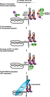
Human telomerase RNP biogenesis. The human TER, hTR, is transcribed by RNA polymerase II as a precursor that cotranscriptionally assembles with the H/ACA protein heterotrimer of dyskerin, NHP2, and NOP10 bound to the H/ACA RNP assembly chaperone NAF1. This process is aided by the dyskerin chaperone SHQ1 and a complex of NUFIP and the helicases RUVBL1 and RUVBL2. NUFIP interacts with NHP2, and the RUVBL1/RUVBL2 heterodimer interacts with dyskerin to promote H/ACA RNP assembly. The hTR precursor transcript is then processed at its 3′ end by an unknown mechanism. The G-quadruplex structure that can form near the 5′ end, represented by a wavy line, is resolved by the helicase DHX36. Then, NAF1 is exchanged for the mature H/ACA RNP protein GAR1, and sTGS1 modifies the 5′ cap to TMG, indicated by a diamond. TCAB1 and TERT bind to hTR in the active telomerase holoenzyme.
Within the hTR H/ACA 3′ hairpin loop, two additional motifs have been defined: the CAB box and the BIO box (Fig. 1). A CAB box is present in each hairpin loop of H/ACA scaRNAs and binds TCAB1/WDR79 (Fig. 5) to promote RNP concentration in Cajal bodies (Richard et al. 2003; Jády et al. 2004; Tycowski et al. 2009; Venteicher et al. 2009). In contrast, the BIO box is not shared by other H/ACA RNAs. Consistent with an hTR-specific requirement for BIO box function, the BIO box cooperates with other hTR-specific 3′ hairpin stem elements to promote mature RNP accumulation in vivo (Fu and Collins 2003; Egan and Collins 2012). BIO box mutant hTR precursor does not escape the site of transcription and is not 3′ end processed (Theimer et al. 2007).
Human telomerase RNP maturation
The hTR precursor is transcribed by RNA polymerase II and exonucleolytically processed at its 3′ end to the boundary of the H/ACA motif (Fig. 5). The presence of a downstream polyadenylation signal or U1 snRNA 3′ box/transcription terminator antagonizes hTR accumulation (Fu and Collins 2003), leaving the actual mechanism of nascent transcript termination and/or endonucleolytic cleavage an open question. The accumulation of mature hTR increases if a self-cleaving ribozyme producing a 2′,3′-cyclic phosphate is inserted immediately downstream from the processed 3′ end (Egan and Collins 2012), suggesting that precursor degradation by 3′ to 5′ exonuclease activity limits hTR accumulation. Unlike all other human H/ACA RNAs, 5′ to 3′ processing of the hTR precursor to the 5′ boundary of the H/ACA domain must be suppressed to retain the template and pseudoknot (Fig. 1).
Many general H/ACA RNP assembly factors are essential for hTR biogenesis (Fig. 5). A preformed H/ACA protein complex of NHP2, NOP10, dyskerin, and the H/ACA RNP assembly factor NAF1 is cotranscriptionally loaded on each hairpin of the nascent H/ACA RNA (Wang and Meier 2004; Darzacq et al. 2006; Richard et al. 2006). While most pseudouridine-guide H/ACA RNAs require the presence of both H/ACA motif hairpins to cooperatively bind the NHP2-NOP10-dyskerin-NAF1 assembly scaffold, hTR exhibits strongly enhanced H/ACA RNP assembly directed by its H/ACA domain 3′ hairpin and dependent on the hTR-specific BIO box (Egan and Collins 2012). The dyskerin chaperone SHQ1 is proposed to function as an RNA mimic, preventing nonspecific RNA binding by dyskerin prior to its assembly into an H/ACA protein core heterotrimer capable of specific H/ACA RNA hairpin binding (Walbott et al. 2011). H/ACA RNP assembly is also aided by a complex of NUFIP, which binds NHP2, and the helicases RUVBL1 and RUVBL2, which bind dyskerin (Boulon et al. 2008; Venteicher et al. 2008). Cellular depletion of any of these chaperone activities reduces mature hTR accumulation in vivo (Hoareau-Aveilla et al. 2006; Fu and Collins 2007; Boulon et al. 2008; Venteicher et al. 2008; Grozdanov et al. 2009).
After transcription, assembly with H/ACA core proteins, and RNA processing (Fig. 5), hTR is routed through Cajal bodies by the transport factors PHAX and Nopp140 (Yang et al. 2000; Boulon et al. 2004). There, the short form of TGS1 (sTGS1) gives hTR its TMG cap (Jády et al. 2004; Fu and Collins 2006; Girard et al. 2008). Cajal bodies are also sites of RNP remodeling, likely including the exchange of the RNP assembly factor NAF1 for the mature RNP component GAR1 (Darzacq et al. 2006). In a manner physically and functionally separable from these steps of RNP biogenesis, the mature RNP also concentrates in Cajal bodies by direct CAB box association with TCAB1/WDR79 (Tycowski et al. 2009; Venteicher et al. 2009). Cellular depletion of TCAB1/WDR79 does not affect hTR accumulation but does reduce RNP association with Cajal bodies and telomeres and results in telomere shortening (Venteicher et al. 2009; Zhong et al. 2011). TCAB1/WDR79 associates with overexpressed hTR partly independent of the CAB box, and, unlike the CAB box, is essential for telomere elongation (Cristofari et al. 2007; Fu and Collins 2007; Egan and Collins 2010; Stern et al. 2012). The significance of TCAB1/WDR79 for telomere maintenance is highlighted by the discovery of TCAB1/WDR79 gene mutations that cause dyskeratosis congenita (Zhong et al. 2011), a human disease of telomerase deficiency (Armanios 2009; Savage and Bertuch 2010).
Human TERT–hTR assembly
TERT is not required for hTR biogenesis or biological accumulation, although it can have a stimulatory effect on the latter when overexpressed (Yi et al. 1999). The TERT TRBD binds the CR4/5 region of hTR, and both the TRBD and TEN domain also bind the template-pseudoknot region (Lai et al. 2001; Robart and Collins 2011). Curiously, at their endogenous levels, for most of the cell cycle hTR concentrates in Cajal bodies, while human TERT concentrates in other nuclear foci (Tomlinson et al. 2006). During S phase, human TERT concentrates in nucleoli and hTR-containing Cajal bodies move to the nucleolar periphery. Only hTR and TERT sites of colocalization, associated with Cajal bodies, are observed to also colocalize with telomeres (Tomlinson et al. 2006). Expression of TERT is required for detectable hTR telomere localization (Tomlinson et al. 2008). Telomeres are not clustered in human cells (Ludérus et al. 1996), and unlike in S. cerevisiae, telomerase appears to associate with most telomeres during a single S phase (Jády et al. 2006; Tomlinson et al. 2006; Zhao et al. 2011). How nuclear subdomains sequester and/or coordinate hTR and TERT complexes remains an open question (Collins 2008). Also open is the possibility of multiple states of telomerase–telomere interaction, for example, distinguishing initial recruitment from productive elongation or accounting for in vivo differences in elongation RAP (Zhao et al. 2011).
As described above for the initial biogenesis of a biologically stable hTR RNP, the subsequent assembly of hTR with TERT is a highly chaperoned process. TERT assembly with hTR is proposed to rely on the protein-folding chaperone HSP90, because the HSP90 inhibitor geldanamycin reduces the production of active human telomerase RNP and induces proteasome-mediated degradation of TERT (Holt et al. 1999; Forsythe et al. 2001; Kim et al. 2005). Expression of a dominant-negative form of the snRNP assembly factor SMN disrupts the localization of TERT in vivo and its assembly with hTR in vitro, suggesting that SMN could play a role in human telomerase RNP catalytic core assembly (Bachand et al. 2002). The DNA-dependent protein kinase-interacting protein KIP could also promote telomerase holoenzyme assembly, since its overexpression increases telomerase activity and telomere length without affecting the levels of TERT mRNA or hTR (Lee et al. 2004). Furthermore, holoenzyme assembly may be regulated by hEST1A/SMG6, an ortholog of yeast Est1 that interacts with hTR and TERT and whose overexpression decreases telomere length (Reichenbach et al. 2003; Snow et al. 2003; Redon et al. 2007).
Bridging the human RNP catalytic core to telomere substrates
Human telomerase recruitment to telomeres requires the TIN2 and TPP1 telomere proteins (Abreu et al. 2010; Tejera et al. 2010), which are anchored to the double-stranded region of the telomere through TRF1 and TRF2 (de Lange 2010; Stewart et al. 2012). The TPP1 OB fold is proposed to interact directly with the TERT TEN domain (Xin et al. 2007; Zaug et al. 2010). TPP1 is also linked to single-stranded telomeric DNA through POT1. A POT1–TPP1 complex stimulates the RAP of a minimal recombinant RNP assembled in rabbit reticulocyte lysate by maintaining association with the product DNA and aiding template translocation (Wang et al. 2007; Latrick and Cech 2010). However, POT1 is not required for telomerase recruitment to telomeres when assayed by ChIP or fluorescence in situ hybridization (Abreu et al. 2010), suggesting that its recruitment role does not require POT1-mediated linkage to single-stranded telomeric DNA.
The human Ku heterodimer has also been suggested to interact with hTR and/or TERT (Chai et al. 2002; Ting et al. 2005), but the in vivo functional significance of the interactions reported to date has not been established. Human Ku70 interacts with TRF2 to localize Ku to telomeres, where it appears to prevent telomere loss (Hsu et al. 1999; Song et al. 2000; Wang et al. 2009). Thus, it is possible that Ku has a role in recruiting human telomerase to telomeres distinct from the roles of Ku in yeast.
COMMON THEMES IN TELOMERASE BIOGENESIS AND REGULATION
Despite remarkably rapid divergence in TER structure and cellular pathways of telomerase RNP maturation, holoenzyme assembly, and recruitment to telomeres, some general features are shared among ciliates, yeasts, and vertebrates. TERs in all three phylogenetic groups recruit proteins prior to TERT binding that remain part of the active holoenzyme and are required for RNA stability in vivo: p65 in ciliates, Sm and Lsm proteins in yeasts, and H/ACA proteins in vertebrates. TERs also contain structurally related template-pseudoknot and STE motifs to mediate their interactions with the TERT TRBD, although the relative affinities of these interactions differ between species. The specificity of TERT–TER interaction in vivo may have remained dependent on the existence of at least two distinct TER–TERT binding interfaces, separated in TER secondary structure but brought together in tertiary structure by prior steps of RNP assembly in vivo. Mechanisms of holoenzyme recruitment to and activation at telomeres also potentially exhibit similarities across phylogenetic groups. The biochemical and regulatory activities of Teb1 and TASC in T. thermophila could parallel the roles of Cdc13, Est1, and Est3 in yeasts and TIN2-bound TPP1 in vertebrates. Future work will be required to further define the precise biological roles of all of the telomerase holoenzyme proteins. Studies of diverse organisms have revealed a great deal of general insight about telomerase biogenesis and regulation, yet much still remains to be understood about the cellular pathways that produce this enzyme and control its activity.
ACKNOWLEDGMENTS
We thank Mary Couvillion for critical reading of this manuscript and members of the Collins lab for helpful discussions. This effort was supported by NIH research grants HL079585 and GM054198 to K.C. and NIH training grant CA009041.
Footnotes
-
↵1 Corresponding author
E-mail kcollins{at}berkeley.edu
-
Article published online ahead of print. Article and publication date are at http://www.rnajournal.org/cgi/doi/10.1261/rna.034629.112.
- Copyright © 2012 RNA Society
REFERENCES
- ↵
- ↵
- ↵
- ↵
- ↵
- ↵
- ↵
- ↵
- ↵
- ↵
- ↵
- ↵
- ↵
- ↵
- ↵
- ↵
- ↵
- ↵
- ↵
- ↵
- ↵
- ↵
- ↵
- ↵
- ↵
- ↵
- ↵
- ↵
- ↵
- ↵
- ↵
- ↵
- ↵
- ↵
- ↵
- ↵
- ↵
- ↵
- ↵
- ↵
- ↵
- ↵
- ↵
- ↵
- ↵
- ↵
- ↵
- ↵
- ↵
- ↵
- ↵
- ↵
- ↵
- ↵
- ↵
- ↵
- ↵
- ↵
- ↵
- ↵
- ↵
- ↵
- ↵
- ↵
- ↵
- ↵
- ↵
- ↵
- ↵
- ↵
- ↵
- ↵
- ↵
- ↵
- ↵
- ↵
- ↵
- ↵
- ↵
- ↵
- ↵
- ↵
- ↵
- ↵
- ↵
- ↵
- ↵
- ↵
- ↵
- ↵
- ↵
- ↵
- ↵
- ↵
- ↵
- ↵
- ↵
- ↵
- ↵
- ↵
- ↵
- ↵
- ↵
- ↵
- ↵
- ↵
- ↵
- ↵
- ↵
- ↵
- ↵
- ↵
- ↵
- ↵
- ↵
- ↵
- ↵
- ↵
- ↵
- ↵
- ↵
- ↵
- ↵
- ↵
- ↵
- ↵
- ↵
- ↵
- ↵
- ↵
- ↵
- ↵
- ↵
- ↵
- ↵
- ↵
- ↵
- ↵
- ↵
- ↵
- ↵
- ↵
- ↵
- ↵
- ↵
- ↵
- ↵
- ↵
- ↵
- ↵
- ↵
- ↵
- ↵
- ↵
- ↵
- ↵
- ↵
- ↵
- ↵
- ↵
- ↵
- ↵
- ↵
- ↵
- ↵
- ↵
- ↵
- ↵
- ↵
- ↵
- ↵




