


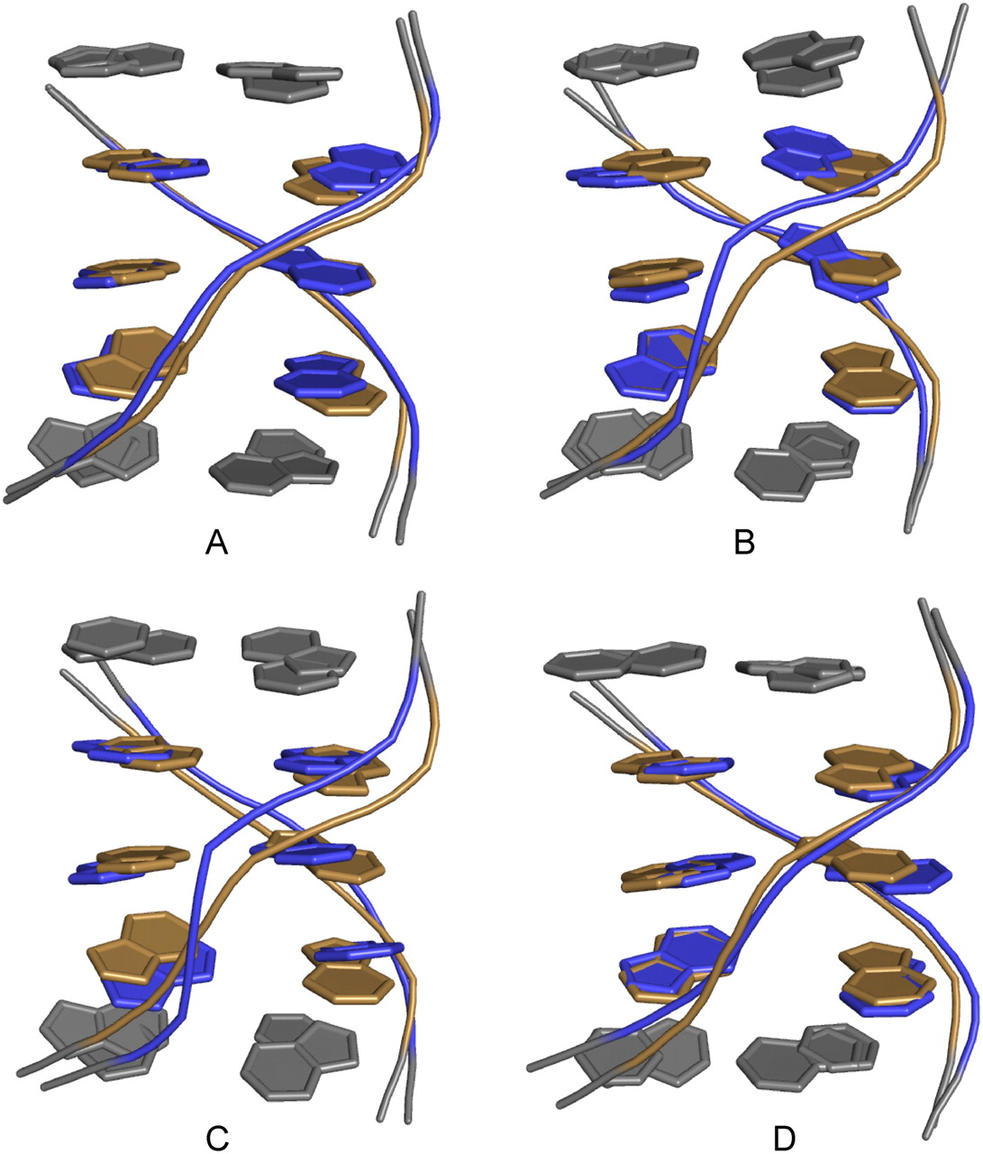
FIGURE 7.
Comparison of B. mori R2 loop (gold) (5′GAAGCC/3′CAGGAG) with other loops (blue) consisting of three sheared purine–purine pairs. (A) 5′CGAAG/3′GAGGU (Chen et al. 2005), (B) 5′UGAAG/3′AAGGU (Ban et al. 2000), (C) 5′UGAGG/3′GAGGU (Schuwirth et al. 2005), and (D) 5′UGAAG/3′GAAGC (Chen et al. 2006). Closing pairs are shown in gray, including the CA pair in the R2 loop. Structures were aligned by pair-fitting of six C1′ atoms in the loop residues.




