NMR structure of a 4 × 4 nucleotide RNA internal loop from an R2 retrotransposon: Identification of a three purine–purine sheared pair motif and comparison to MC-SYM predictions

(Downloading may take up to 30 seconds. If the slide opens in your browser, select File -> Save As to save it.)

Click on image to view larger version.

FIGURE 3.
FIGURE 3.

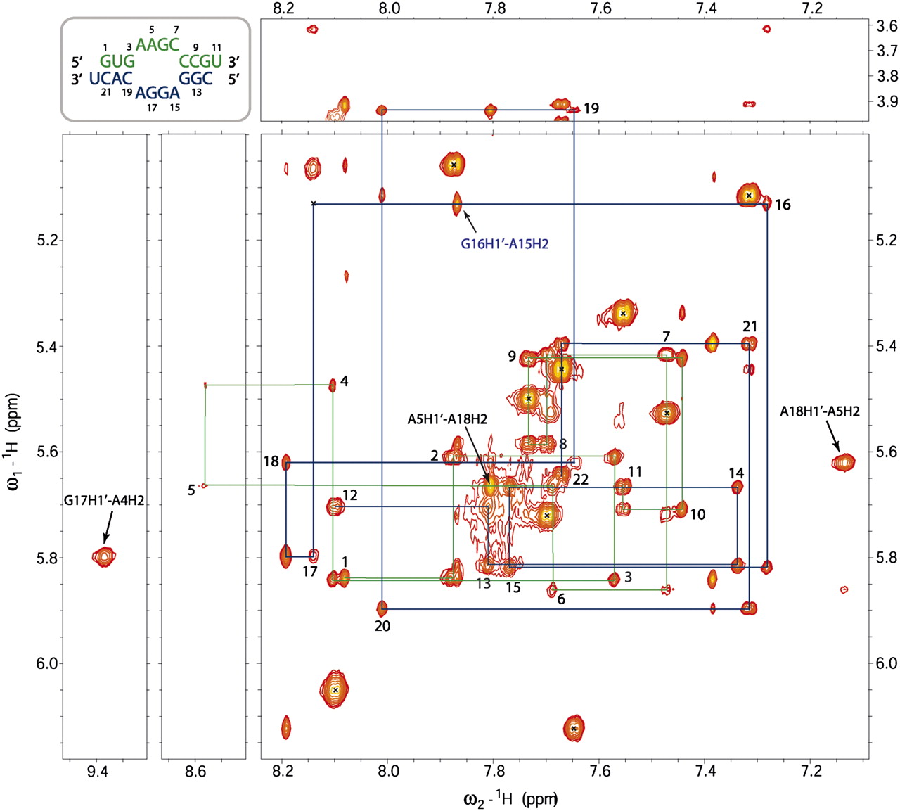
2D NOESY spectrum of the R2 internal loop in 100% D2O at 20°C showing the H8/H6/H2H1′/H5 region. The mixing time is 400 msec. C and U H5–H6 NOEs are indicated by crosses. Important cross-strand NOEs involving adenine H2 protons are indicated in black.

This Article

  1. RNA 17: 1664-1677

