


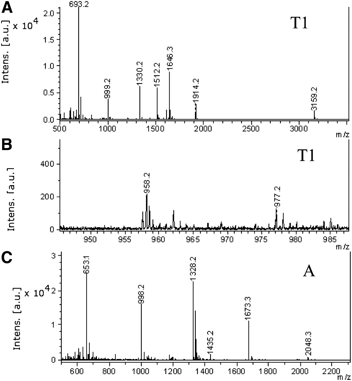
FIGURE 2.
MALDI analyses of RNase digests of tRNAArg. (A) T1 digest, major peaks; (B) detail of 860–1000 m/z region of T1 digest, showing presence of minor peak at 977.2 m/z, corresponding to CD17G 3′P; (C) A digest, major peaks.
(Downloading may take up to 30 seconds. If the slide opens in your browser, select File -> Save As to save it.)
Click on image to view larger version.
MALDI analyses of RNase digests of tRNAArg. (A) T1 digest, major peaks; (B) detail of 860–1000 m/z region of T1 digest, showing presence of minor peak at 977.2 m/z, corresponding to CD17G 3′P; (C) A digest, major peaks.
 CiteULike
 Delicious
 Digg
Digg Facebook
 Reddit
 Twitter
Molecular Cloning
The New Edition
Buy Now and Save 30%
Paperback only
(limited time offer)

|
Molecular Cloning: A Laboratory Manual (Fourth Edition) |
