


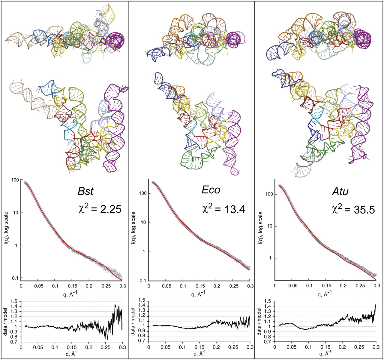
FIGURE 3.
(Top) All-atom models of the RNase P RNA variants. RNA models were generated in silico by taking into account available crystallographic and phylogenetic information. Elements of the structure are colored according to Figure 1. (Middle) Agreement between theoretical scattering profiles calculated from the models (red line) and experimental SAXS data (open circles). (Bottom) Distribution of the residuals (Idata/Imodel) vs. q.




