


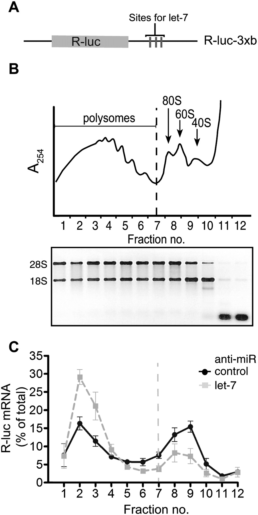
Manipulation of let-7 activity by anti-miR transfection. HeLa cell lysates were prepared 9 h after transfection with R-luc-3xB reporter and anti-miR against let-7 or control anti-miR, followed by density gradient ultracentrifugation and RNA analysis. (A) Schematic of the Renilla luciferase R-luc-3xB reporter. (B) Representative absorbance trace at 254 nm is shown at the top to locate positions of polysomes, ribosomes (80S), and subunits (60S, 40S). Gel analysis of gradient fraction RNA integrity is shown below. (C) Gradient distribution of R-luc-3xB mRNA after anti-let-7 (gray squares) or control anti-miR transfection (black circles) was measured by qRT–PCR. R-luc-3xB mRNA abundance in each fraction is expressed as a percentage of the total R-luc-3xB mRNA in the gradient (see Supplemental Fig. S1 for normalization strategy). Dotted line represents border between polysomal and subpolysomal complexes. Data are an average of three experiments, and bars represent standard error.




